2. Early Onset Scoliosis
Learning Objectives Concerning: Etiology, Classification, and Natural History
- Learner will be able to define early onset scoliosis (EOS).
- Learner will be able to describe the prevalence and etiologies of EOS.
- Learner will be able to describe associated conditions, and evaluation of the patient presenting with EOS.
- Learner will be able to discuss the current EOS classification systems (including congenital scoliosis).
- Learner will be able to describe the natural history of untreated EOS and relationship of EOS to pulmonary function.
- Learner will be able to describe the growth of the thoracic spine and thorax and the development of thoracic insufficiency syndrome.
Learning Objectives Defining and Identifying: Clinical and Radiological Parameters; Patient's HRQoL, and Guiding the Treatment Decision Making Process
- Learner will be able to interpret clinical and radiological parameters in EOS.
- Learner will be able to estimate the patient’s risk of progression based on the natural history of the deformity.
- Learner will be able to list the indications, risks and benefits for non-operative treatment vs. surgical intervention.
- Learner will describe the use of HRQoL data and appropriate outcome measures.
Learning objectives concerning: defining, formulating and recommending treatment options, considering safety, and predicting outcomes and potential complications
- Learner will be able to describe the technique of corrective cast application for EOS.
- Learner will be able to describe the use of halo gravity traction in the treatment of EOS including complications.
- Learner will be able to list the risk, benefits and complications of various growth friendly implant and correction strategies (e.g. distraction based, compression based, growth guided).
- Learner will be able to describe the indications and complications of osteotomy and fusion based treatment including hemivertebrectomy in the treatment of EOS.
- Learner will be able to describe the neuromonitoring indications and unique features of patients with EOS.
- Learner will be able to select best treatment options considering capacity for appropriate follow-up for each patient throughout remaining growth.
- Learner will be able to select fusion levels using clinical and radiographic parameters to optimize spinal balance and motion preservation.
- Learner will describe use of measures to improve safety in EOS surgery and minimize perioperative complications.
- Learner will be able to discuss complication management for the treatment options in EOS.
Author: Colin Nnadi, FRCS (Orth)
Key Points
- Early onset neuromuscular scoliosis incorporates multiple aetiologies
- Due to better understanding of genetics and better study design, historical evidence of cerebral palsy may not accurately reflect underlying aetiology
- New disease modifying therapies mean surgery may be delayed for several years
- An understanding of natural history is crucial in appropriate treatment strategies
- Several growth-friendly options available but definitive fusion may be only option in certain cases
Abstract
Early onset scoliosis (EOS) is defined as spinal deformity of over 100 presenting before the age of 10 years. EOS is a heterogenous group consisting of congenital, syndromic, neuromuscular and idiopathic etiologies. Although a lot has been written about EOS there remains a paucity of literature on the individual aetiologies because of the small numbers involved. Most of the natural history studies are focused on the infantile idiopathic group and in the case of early onset neuromuscular scoliosis focus on treatment and its effects but do not delve into the natural history or different types. Some of the earlier European studies seemed to reinforce the view EOS was primarily a European problem (12.8%) compared to 0.25% in North America (1). In this article we summarise some of the more common presentations of early onset neuromuscular scoliosis and discuss the role of non-operative and surgical treatment options.
Key words – early onset, neuromuscular scoliosis, heterogenous, natural history.
Neuromuscular scoliosis
Neuromuscular scoliosis is spread across a large spectrum of pathologies the manifestations of which will evolve with growth over the natural history. Each of these pathologies is unique in presentation and its response to treatment. Treatment aims to achieve the following:
- Optimal spinal growth
- Prevent spinal deformity progression
- Optimization thoracic and lung
development - Maintain ambulation if possible
- Improve seating comfort
- Improve general well being
Neuromuscular EOS (NM-EOS) is a heterogenous group within the EOS population. Clinical series quote 20-40% of EOS cohorts. Familiarity with the different etiologies and subsequent comorbidities and prognosis is essential in planning treatment.
Treatment
Treatment which can be non-operative or surgical does provide improvements in quality of life (QOL) parameters. Surgical treatment has led to improved spinal deformity correction with modern techniques (2, 3, 4,5). High parent/caregiver and patient satisfaction rates have been reported following surgery. Watanabe et al performed a retrospective review of 84 patients/families of spastic cerebral palsy (CP) patients undergoing spinal fusion. The mean age at the time of surgery was 15.8 years (9 to 26 years). Average follow-up was 6.2 years. They reported overall satisfaction rate of 92%. Ninety three percent reported improvement with sitting balance, 94% with cosmesis, and 71% in patient’s quality of life. Functional improvements were however restricted. Satisfaction rates were lower in patients with increased complication rates and residual deformity post-operatively or in those who were less disabled pre-operatively (6).
Despite the high satisfaction scores complication rates for surgery remain high. Rumalla et al reported on neuromuscular scoliosis treated between 2002 and 2011. They examined 2154 cases. Overall complication rate was 40%. (7). Brandon et al (8) reported on a 30-year experience at a single institution on surgical site infections after posterior spinal fusion for neuromuscular scoliosis. They looked at 428 patients with average follow-up of 4.9 years. Mean Cobb angle was 74 degrees. Deep infection rate was 10.3%. More than half of these infections occurred within 3 months of surgery. 45% were polymicrobial and up to 60% were gram positive. Risk factors identified were diagnosis of spina bifida, BMI <25kg/m2 , incontinence, non-ambulation, Albumin <3.5g/dL, Zinc >60 ASA 3 grade and pelvic fixation. Other authors (9) report complication rates with a prevalence of 28% and also identify pre-operative curve magnitude >60 degrees and non-ambulatory status as risk factors. Yaszay et al (10) reported on 257 patients with follow up > 2years. They found a post-operative major complication rate of 36% and spine related re-operation rate of 14%. Treating surgeons should therefore bear in mind that complication rates with surgical management of neuromuscular scoliosis are not insignificant. It is important to ensure patients are adequately worked up for surgery. Informed consent highlighting areas where improvement in QOL is likely to occur post-operatively will also help to manage parents/care-givers outcome expectations.
Current Surgical Techniques
The outcomes for early spinal fusion in EOS are unclear but a retrospective study by Sitoula et al (11) looked at 33 children who underwent spine fusion with unit-rod instrumentation between 1989 and 2006 for CP neuromuscular scoliosis, aged below 10 years at spine fusion, and with follow-up >5 years. They found that spine fusion was associated with minimal short-term and long-term morbidity, but there was 28% mortality at 10 years of follow-up and 50% predicted mortality at 15 years. Modern practice on treatment of neuromuscular scoliosis has now shifted to the use of Growing Rod technologies with the aim of preserving growth and development similar to the idiopathic group where possible. In certain cases the risks associated with this approach outweigh the benefits and early spine fusion may be the only option. Most growth-friendly constructs for neuromuscular EOS will fall into the distraction based or growth guided categories. The surgical objectives are as follows:
- Optimise spine and lung growth where possible
- Balance spine
- Correct pelvic obliquity
- Improve sitting posture
- Minimise complications
Surgical options available:
- Distraction based – Traditional growing rods (TGR), Magnetically controlled growing rods (MCGR), VEPTR
- Guided growth – Shilla, Luque trolley, modified Luque trolley, Unit Rod, Sub-laminar bands
Different surgical constructs as stated above are available for treatment of progressive neuromuscular EOS and choice will often fall to the treating surgeon depending on preference. Each system has its pros and cons. Numerous authors have published their experiences. McElroy et al performed a retrospective evaluation of 27 children with TGR through a posterior only approach. 15 of the constructs extended to the pelvis. They reported that TGRs were effective and constructs extending to the pelvis controlled pelvic obliquity more effectively. 30% of patients experienced deep wound infection (12). Tsirikos et al in 2008 reported on 287 children with unit rod instrumentation in an observational study. 241/287 had >2year follow up. Scoliosis correction was from mean Cobb angle of 76 degrees to 25 degrees (68%) and pelvic obliquity correction was from 17 degrees to 5 degrees (71%)(13). Lonstein et al in 2012 reported on the use of the Luque Galveston technique in a retrospective analysis of 93 patients. They concluded that it was a safe and effective procedure (14). Canavese et al (15) reported 49% correction in neuromuscular curves and found that a physiological kyphosis could be achieved in 88% of the patients using sublaminar bands. Gersoff et al (16) in their review of 33 patients followed up over 40 months reported no rod failures or pseudoarthrosis. They concluded that the Luque technique was effective.
More recent technologies such as MCGR and Growth guidance systems have been shown to be effective in the surgical treatment of EOS. Haapala et al (17) compared the health-related quality of life (HRQoL) and surgical outcomes of Shilla growth guidance and magnetically controlled growing rod (MCGR) treatment in patients with syndromic and neuromuscular early-onset scoliosis. They concluded that both systems offered similar correction of spinal deformity with equal number of complications but the Shilla system had less surgical procedures in patients treated for EOS when compared with MCGR instrumentation. MCGR resulted in slightly better spinal growth during the distraction period. There were no differences in the QOL measurements. The drawback was the limited numbers in this series. (17). In another study Swarup et al (18) looked at patients with spinal muscular atrophy treated surgically with growth friendly devices and the effect of these constructs and the addition of chest wall support (CWS) on spinal deformity, thorax morphology and pulmonary outcomes. There were 66 patients – 25% MCGR, 73% VEPTR and 2% unknown. They concluded that the constructs did improve the spinal deformity but those treated with CWS had better improvements in thoracic morphology compared to those without.
Despite the obvious benefits of surgery there are challenges associated with achieving good outcomes. There is an increased risk of blood loss therefore tranexamic acid and use of cell salvage is standard. In the author’s (CN) institution >40% blood loss is an indication to abort the procedure. The decision on whether or not to use pelvic fixation in ambulatory patients is complex as there is risk of impairment of mobility, which is distressing for patients and care-givers due to loss of independence. Technically, implants can be prominent with resultant skin breakdown. The surgical approach is often extensive even with growth friendly techniques, which can give rise to healing complications particularly in the distal aspect of the wound due to the risk of faecal or urinary contamination. A modified Wiltse approach (19) has been described to minimise the extensive tissue disruption normally associated with surgery through a conventional approach. The authors reported lower blood loss and comparable curve correction to the traditional midline approach.
Controversies in Surgical Management of Neuromuscular Scoliosis
Intra-operative monitoring
There is controversy around the usefulness of IOM in neuromuscular patients under-going surgery. Dulfer et al compared 15 patients with DMD and 15 patients with AIS to investigate the feasibility of IONM and cortical excitability. They concluded that IOM seems a feasible and valuable neurophysiological tool during scoliosis correction surgery in patients with DMD (20). Ashkenaze et al (21) reported on their series of 101 patients. They found in 28% a reliable tracing could not be obtained. Noordeen et al (22) in a retrospective review of the role of monitoring of somatosensory spinal evoked potentials (SSEP) in 99 patients with neuromuscular scoliosis showed that SSEP monitoring was useful and that a 50% decrease in the amplitude of the trace optimised both sensitivity and specificity.
Depending on aetiology and patient base line status, intra-operative neural monitoring can be considered. Sometimes due to disease severity, monitoring may not be possible and this should be discussed in advance with the patients or care-givers.
L5 or Pelvis?
The decision on where to stop the instrumentation in Neuromuscular scoliosis is multi-factorial. The magnitude of the deformity and the presence of pelvic obliquity are important but the functional status of the patient is as well. Patients with a higher GMFCS (IV or V) grade are more likely to require fixation to the pelvis compared to those with lower grades. Tondevold et al (23) in a retrospective study compared fixation to L5 and to the pelvis in non-ambulatory patients. They found that patients undergoing pelvic fixation had a greater correction of Cobb angle and pelvic obliquity with no difference in rate of complications between the 2 groups. Different techniques include Galveston technique, iliac screws, Maximum Width Construct, Dunn McCarthy, Sacral Alar Iliac (SAI) and spinopelvic trans-iliac fixation system.
Early Spine Fusion
In a landmark paper Karol (48) reviewed pulmonary function at intermediate-term follow-up in 28 patients with scoliosis who underwent thoracic fusion before the age of nine years. Forced vital capacity was <50% of normal in 12/28 patients, and two required respiratory support. Sitoula et al (49) reported on 10-year outcomes of early fusion in children with cerebral palsy. There was minimal short and long-term morbidity but 28% mortality at 10 years and 50% predicted mortality at 15 years. The decision to offer early fusion as opposed to growth friendly surgery is very much dependent on patient circumstances, natural history of the condition and surgeon experience.
Non-Operative Management
The aim of non-operative management is to control curve progression and improve seating comfort. The mainstay of non-operative treatment is bracing, Wheelchair support and therapies. Recent reports (24,25) indicate that bracing does improve sitting balance and trunk support and by extrapolation leads to better neck and head control. Braces can be used to assist with wheelchair seating as well. It is usually an interim support measure in this group of patients before surgical intervention.
Wheelchair seating in non-ambulatory patients optimises sitting comfort and improves function (26). A 3-point system of lateral support pads has been shown to offer a more symmetrical trunk posture and correction of curve angles by 35% in non-ambulatory CP patients with scoliosis (27). Other non-operative interventions include Botulinum toxin injections and intrathecal baclofen infusions which have both been shown to be effective in the management of neuromuscular scoliosis (28,29).
Different Etiologies in Neuromuscular Scoliosis
Cerebral Palsy
Cerebral palsy is a non-progressive neurological disorder. The prevalence of scoliosis in patients with cerebral palsy (CP) has been reported at up to 72% (30,31, 32,33). The Southern Swedish Cerebral Palsy Registry in 2018 reviewed patients with cerebral palsy and reported that individuals who function at higher Gross Motor Function Classification System (GMFCS) levels are most at risk of developing scoliosis. (34). However, there should be a balance between the benefits of spinal surgery in children with cerebral palsy and the well-known higher risks of postoperative complications, which can be multi-systemic and include but are not exclusive to wound infections, skin breakdown, respiratory/cardiovascular compromise as well as gastrointestinal complications (35,36). There are challenges with interpretation of the natural history of scoliosis in cerebral palsy due paucity of high quality data but available evidence indicates that progression continues even after skeletal maturity. Patients with poor functional status are more likely to progress and should be considered for surgery at an appropriate time.
Friedreichs Ataxia
Friedreich’s ataxia (FA) is a progressive degenerative autosomal recessive condition caused by a deficiency of frataxin. It is a multi-systemic disorder which affects the central and peripheral nervous systems, heart, skeleton, and endocrine pancreas.
Before genetic confirmation was possible patients were diagnosed clinically and described by their neurological phenotype and presentation as, typical <20 years of age, or atypical ( >20 years). Labelle et al (37) reported on 56 patients with typical FA. The group were followed up for an average of 9 years (range 1 to 16 years). A scoliosis of more than 10 degrees was found in all patients and was associated with a hyperkyphosis in 66 per cent. Both sexes were equally affected. Fifty-seven per cent of the curves were double thoracic and lumbar; 14 per cent were thoracolumbar; 7 per cent, double thoracic and thoracolumbar; 7 per cent, thoracic; 4 per cent, lumbar; and 11 per cent, multiple small curves. The group was divided into 3 roughly equal groups with respect to their spinal profile. 30% had a clearly relentless progression of scoliosis to over 60 degrees Cobb angle. The second 30% had a scoliosis; however, this had stayed under 40 degrees without progression. The final 30% had a scoliosis that was under 40 degrees. This disputes an earlier assumption that FA is relentlessly progressive. LaPaen et al (38) investigated predictors for the clinical course of a child with FA. They found a correlation between genotype modifiers and the age at which a child had to use a wheelchair. The natural history of the spine in FA is likely to be of an idiopathic/ syndromic nature and is not always aggressive. This may infer that fixation to the pelvis is not always necessary.
Spinal Muscular Atrophy (SMA)
SMA is a hereditary autosomal recessive condition affecting the second motor neuron, causing progressive muscle atrophy and weakness due to decreased expression of the survival motor neuron. There are different sub-types which reflect survival rates. It is characterized by progressive scoliosis and pelvic obliquity. Disease severity stratification correlates with age at presentation and developmental milestones achieved. The prognosis is dependent on type and this may influence surgical or non-surgical decision making (39).
In 2016 Nusinersen (50) was approved for all types of SMA. It is a splicing modifier that enhances the expression of survival motor neuron and requires intrathecal administration. It has changed the natural history of the condition and previously scoliosis surgery was an exclusion factor for treatment. The bony fusion mass was viewed as an obstruction to introducing the needle for Nursinersen administration. Weinstein et al have previously described a technique for securing intrathecal access (51).
Duchenne Muscular Dystrophy
Duchenne muscular dystrophy (DMD) is an X-linked recessive neuromuscular disorder caused by mutations in the dystrophin gene that result in absent or insufficient functional dystrophin (40). There is a gradual and progressive decline in muscle strength correlating with development of scoliosis. There is a relentless progression of scoliosis in boys with DMD after loss of ambulation (41). In recent years there have been new emerging developments which have altered the natural history of scoliosis in the DMD affected patients. King et al in 2007 (42) in a retrospective study of 143 boys and young men recruited from 2000 to 2003, reported on the orthopaedic outcomes of daily corticosteroid use in DMD. The treated group (53%) had steroids for more than a year. The untreated group (47%) had taken steroids for less than 6 months or not at all. The development of scoliosis was only reported after 9 years of age. In another prospective cohort from Toronto (43). Alman reported the use of steroids and development of scoliosis in DMD. Two groups of ambulant boys aged 7 to 10 years old, were enrolled in a non-randomized comparative study to determine the effect of deflazacort (a derivative of prednisone) on muscle strength and pulmonary function. Parental choice dictated the use of steroids. 30 patients were treated with deflazacort (treatment group), and 24 were not (control group). Follow up was to a mean age of 16 years (15-18) in both groups. The mean follow-up was 7.3 years (5-8).
Curve progression of ≥20° developed during the follow-up period in 16/24 (67%) in the control group but in only 5/30 (17%) in the treatment group. 15/24 patients in the control group had spine surgery, at a mean age of 13 years, (11-17); at time of surgery, the mean Cobb angle was 35° (20° - 60°). On the other hand only 5/30 patients in the treatment group underwent spine surgery, at mean age of 15 years (11-17). Biggar in his series noted that by 18 years of age 30/34 (90%) boys not treated had developed a spinal curve of 20 degrees and had surgery, compared to 4/40 (10%) treated boys who had a curve above 20 degrees and who had surgery. Whilst these studies seem to show that steroid treatment slows the development of scoliosis, it is unclear if the decrease in scoliosis seen at the time was due to increased age at loss of ambulation or the lack of decrease in strength that is afforded by use of steroids. In another study Lebel at el (44) reported on 54 boys with DMD in a non-randomised comparative study. They concluded that the long-term use of the glucocorticoid resulted in a substantial decreased need for spinal surgery to treat scoliosis.
In addition to the beneficial effects of steroid therapy, maintaining ambulation with rehabilitation into Knee Ankle Foot Orthoses (KAFO’s) at the beginning of loss of ambulation has been found to prolong time that the boys are ambulant by about 2 years (45) and is standard practice. It is suggested that maintaining this standing and walking ability may help in preventing the rapid progression of scoliosis in DMD (46). In another study Kinali et al (47) looked at 123 boys who were known to their service, and who in 2007 were 17 and over. 78% had been rehabilitated into KAFO’s at loss of ambulation and 30% had been treated with steroids, with a varied length of treatment, range 2 months to 9 years, median 1 year. The authors were able to demonstrate a strong association both between later age of loss of ambulation and length of steroid use in delaying onset of scoliosis, with older age at loss of ambulation being associated with lesser curves.
Summary
The natural history of early onset neuromuscular scoliosis is dependent on aetiology. An insight into likelihood of progression, rate of progression and life expectancy facilitate decision making on treatment.
In the modern era disease modifying therapies have tempered the need for immediate surgical management. Advanced bracing techniques and wheelchair design imply that surgical delays of several years is the new norm. An improved awareness of the diversity of aetiology has allowed us a better appreciation of conditions such as cerebral palsy. Previously, historical evidence of the natural history relied heavily on retrospective series from adults in residential settings. The diagnosis of cerebral palsy has now changed from one of a presumed diagnosis in a physically or learning-disabled child, to one where evidence of a non-progressive lesion in a developing brain can be identified. Advanced imaging techniques and genetic testing means that a previously labelled cerebral palsy cohort would now be classified as a specific or syndromic condition. Better medical management means these patients are now living longer and decisions on surgery should be tempered with potential effects on long-term morbidity and mortality.
References
1. McMaster MJ. Infantile idiopathic scoliosis: can it be prevented? J Bone Joint Surg Br. 1983;65:612-7, Wynne-Davies R. Familial (idiopathic) scoliosis. A family survey. J Bone Joint Surg Br. 1968;50:24-30
2. Allen BL Jr, Ferguson RL. L-rod instrumentation for scoliosis in cerebral palsy. J Pediatr Orthop 1982;2:87–96, Bell DF, Moseley CF, Koreska J. Unit rod segmental spinal instrumentation in the management of patients with progressive neuromuscular spinal deformity. Spine 1989;14:1301–7
3. Bonnett C, Brown JC, Grow T. Thoracolumbar scoliosis in cerebral palsy: results of surgical treatment. J Bone Joint Surg Am 1976;58:328–36
4. Brown JC, Swank S, Specht L. Combined anterior and posterior spine fusion in cerebral palsy. Spine 1982;7:570–3
5. Lonstein JE, Akbarnia B. Operative treatment of spinal deformities in patients with cerebral palsy or mental retardation: an analysis of one hundred and seven cases. J Bone Joint Surg Am 1983;65:43–55
6. Is Spine Deformity Surgery in Patients With Spastic Cerebral Palsy Truly Beneficial?A Patient/Parent Evaluation Kota Watanabe, MD, Lawrence G. Lenke, MD, Michael D. Daubs, MD, Kei Watanabe, MD, Keith H. Bridwell, MD, Georgia Stobbs, RN,and Marsha Hensley, RN, BS
7. Rumalla K, Yarbrough CK, Pugey AJ. Spinal fusion for pediatric neuromuscular scoliosis: national trends, complications and in hospital outcomes. J Neurosurg Spine 2016;25:500–8
8. Ramo, Brandon A. MD; Roberts, David W. MD; Tuason, Dominick MD; McClung, Anna RN, BSN; Paraison, Lauren E. BA; Moore, Harold G. IV; Sucato, Daniel J. MD Surgical Site Infections After Posterior Spinal Fusion for Neuromuscular Scoliosis, The Journal of Bone and Joint Surgery: December 17, 2014 - Volume 96 - Issue 24 - p 2038-2048
9. Master, Daniel L. MD; Son-Hing, Jochen P. MD, FRCS (C); Poe-Kochert, Connie RN, CNP; Armstrong, Douglas G. MD, FRCS (C); Thompson, George H. MD Risk Factors for Major Complications After Surgery for Neuromuscular Scoliosis, Spine: April 01, 2011 Volume 36 - Issue 7 - p 564-571
10. Yaszay B, Bartley CE, Sponseller PD, Abel M, Cahill PJ, Shah SA, Miyanji F, Samdani AF, Daquino C, Newton PO. Major complications following surgical correction of spine deformity in 257 patients with cerebral palsy. Spine Deform. 2020 Dec;8(6):1305-1312.
11. The Long-term Outcome of Early Spine Fusion for Scoliosis in Children With Cerebral Palsy. Sitoula, Prakash MD; Holmes, Laurens Jr PhD, DrPH; Sees, Julieanne DO; Rogers, Kenneth PhD, ATC; Dabney, Kirk MD; Miller, Freeman MD.
12. McElroy MJ, Sponseller PD, Dattilo JR, Thompson GH, Akbarnia BA, Shah SA, Snyder BD; Growing Spine Study Group. Growing rods for the treatment of scoliosis in children with cerebral palsy: a critical assessment. Spine (Phila Pa 1976). 2012 Nov 15;37(24):E1504-10.
13. Tsirikos AI, Lipton G, Chang WN, Dabney KW, Miller F. Surgical correction of scoliosis in pediatric patients with cerebral palsy using the unit rod instrumentation. Spine (Phila Pa 1976). 2008 May 1;33(10):1133-40
14. Lonstein JE, Koop SE, Novachek TF, Perra JH. Results and complications after spinal fusion for neuromuscular scoliosis in cerebral palsy and static encephalopathy using luque galveston instrumentation: experience in 93 patients. Spine (Phila Pa 1976). 2012 Apr 1;37(7):583-91
15. Canavese F, Samba A. Sublaminar polyester bands for the correction of idiopathic and neuromuscular scoliosis. Ann Transl Med. 2020;8(2):32
16. Gersoff WK, Renshaw TS. The treatment of scoliosis in cerebral palsy by posterior spinal fusion with Luque-rod segmental instrumentation. J Bone Joint Surg Am. 1988 Jan;70(1):41-4.
17. Haapala H, Saarinen AJ, Salonen A, Helenius I. Shilla Growth Guidance Compared with Magnetically Controlled Growing Rods in the Treatment of Neuromuscular and Syndromic Early-onset Scoliosis. Spine (Phila Pa 1976). 2020 Dec 1;45(23):E1604-E1614
18. Swarup I, MacAlpine EM, Mayer OH, Lark RK, Smith JT, Vitale MG, Flynn JM, Anari JB; Pediatric Spine Study Group, Cahill PJ. Impact of growth friendly interventions on spine and pulmonary outcomes of patients with spinal muscular atrophy. Eur Spine J. 2021 Mar;30(3):768-774
19. The Value of a Modified Wiltse Approach for Deformity Correction in Neuromuscular Scoliosis, Kieser et al., Int J spine Surg, 2020
20. Dulfer, Sebastiaan & Potgieser, A. & Groen, Rob & Drost, Gea & Lange, Fiete & Wapstra, F. & Faber, C & Valk, J. & Absalom, Anthony. (2020). Intraoperative neurophysiological monitoring during scoliosis surgery in patients with Duchenne muscular dystrophy. European Spine Journal. 29. 1-7. 10.1007
21. Ashkenaze D, Mudiyam R, Boachie-Adjei O, Gilbert C. Efficacy of spinal cord monitoring in neuromuscular scoliosis. Spine (Phila Pa 1976). 1993 Sep 15;18(12):1627-33
22. Noordeen MH, Lee J, Gibbons CE, Taylor BA, Bentley G. Spinal cord monitoring in operations for neuromuscular scoliosis. J Bone Joint Surg Br. 1997 Jan;79(1):53-7
23. Tøndevold N, Lastikka M, Andersen T, Gehrchen M, Helenius I. Should instrumented spinal fusion in nonambulatory children with neuromuscular scoliosis be extended to L5 or the pelvis? Bone Joint J. 2020 Feb;102-B(2):261-267. doi: 10.1302/0301-620X.102B2.BJJ-2019-0772.R2
24. Terjesen T, Lange JE, Steen H. Treatment of scoliosis with spinal bracing in quadriplegic cerebral palsy. Dev Med Child Neurol 2000;42:448-54. 10.1017
25. Miller A, Temple T, Miller F. Impact of orthoses on the rate of scoliosis progression in children with cerebral palsy. J Pediatr Orthop 1996;16:332-5. 10.1097
26. Angsupaisal M, Maathuis CG, Hadders-Algra M. Adaptive seating systems in children with severe cerebral palsy across International Classification of Functioning, Disability and Health for Children and Youth version domains: a systematic review. Dev Med Child Neurol. 2015 Oct;57(10):919-30
27. Holmes KJ, Michael SM, Thorpe SL, Solomonidis SE. Management of scoliosis with special seating for the non-ambulant spastic cerebral palsy population--a biomechanical study. Clin Biomech (Bristol, Avon). 2003 Jul;18(6):480-7
28. Hoare BJ, Wallen MA, Imms C, Villanueva E, Rawicki HB, Carey L. Botulinum toxin A as an adjunct to treatment in the management of the upper limb in children with spastic cerebral palsy (UPDATE). Cochrane Database Syst Rev. 2010 Jan 20;2010(1):CD003469
29. Albright AL, Cervi A, Singletary J. Intrathecal baclofen for spasticity in cerebral palsy. JAMA. 1991 Mar 20;265(11):1418-22
30. Balmer GA, MacEwen GD. The incidence and treatment of scoliosis in cerebral palsy. J Bone Joint Surg Br 1970;52:134–7
31. Edebol-Tysk K. Epidemiology of spastic tetraplegic cerebral palsy in Sweden. Part I: impairments and disabilities. Neuropediatrics 1989;20:41–5
32. Madigan RR, Wallace SL. Scoliosis in the institutionalized cerebral palsy population. Spine 1981;6:583–90
33. Rosenthal RK, Levine DB, McCarver CL. The occurrence of scoliosis in cerebral palsy. Dev Med Child Neurol 1974;16:664–7
34. Hagglund G, Pettersson K, Czuba T, Persson-Bunke M, Rodby-Bousquet E. Incidence of scoliosis in cerebral palsy. Acta Orthop. 2018;89:443-447
35. Nishnianidze T, Bayhan IA, Abousamra O, Sees J, Rogers KJ, Dabney KW, Miller F. Factors predicting postoperative complications following spinal fusions in children with cerebral palsy scoliosis. Eur Spine J. 2016;25:627-634
36. Samdani AF, Belin EJ, Bennett JT, Miyanji F, Pahys JM, Shah, SA, Newton PO, Betz RR, Cahill PJ, Sponseller PD. Major perioperative complications after spine surgery in patients with cerebral palsy: assessment of risk factors. Eur Spine J. 2016;25:795-800
37. Labelle H, Tohmé S, Duhaime M, Allard P. Natural history of scoliosis in Friedreich's ataxia. J Bone Joint Surg Am. 1986 Apr;68(4):564-72
38. La Pean A, Jeffries N, Grow C, Ravina B, Di Prospero NA. Predictors of progression in patients with Friedreich ataxia. Mov Disord. 2008;23(14):2026-2032.
39. Natural course of scoliosis in proximal spinal muscular atrophy type II and IIIa: descriptive clinical study with retrospective data collection of 126 patients, Fujak et al, BMC Musculoskeletal Disorders, 2013
40. Birnkrant DJ, Bushby K, Bann CM, et al. Diagnosis and management of Duchenne muscular dystrophy, part 1: diagnosis, and neuromuscular, rehabilitation, endocrine, and gastrointestinal and nutritional management [published correction appears in Lancet Neurol. 2018 Apr 4;:]. Lancet Neurol. 2018;17(3):251-267
41. Wilkins KE, Gibson DA. The patterns of spinal deformity in Duchenne muscular dystrophy. J Bone Joint Surg Am 1976;58(1)
42. Orthopedic outcomes of long-term daily corticosteroid treatment inDuchenne muscular dystrophy W.M. King, R. Ruttencutter, H.N. Nagaraja, V. Matkovic, J. Landoll, C. Hoyle, J. R. Mendell, J. T. Kissel Neurology May 2007, 68 (19) 1607 1613
43. W.D. Biggar, V.A. Harris, L. Eliasoph, B. Alman,Long-term benefits of deflazacort treatment for boys with Duchenne muscular dystrophy in their second decade,Neuromuscular Disorders,Volume 16, Issue 4, 2006. Pages 249-255,ISSN 0960-8966
44. Lebel DE, Corston JA, McAdam LC, Biggar WD, Alman BA. Glucocorticoid treatment for the prevention of scoliosis in children with Duchenne muscular dystrophy: long-term follow-up. J Bone Joint Surg Am. 2013 Jun 19;95(12):1057-61
45. Bakker JP, de Groot IJ, Beckerman H, de Jong BA, Lankhorst GJ. The effects of knee-ankle-foot orthoses in the treatment of Duchenne muscular dystrophy: review of the literature. Clin Rehabil. 2000 Aug;14(4):343-59. doi: 10.1191/0269215500cr319oa. PMID: 10945419.
46. Rodillo EB, Fernandez-Bermejo E, Heckmatt JZ, Dubowitz V. Prevention of rapidly progressive scoliosis in Duchenne muscular dystrophy by prolongation of walking with orthoses. J Child Neurol. 1988 Oct;3(4):269-74.
47. Kinali M, Main M, Eliahoo J, Messina S, Knight RK, Lehovsky J, Edge G, Mercuri E, Manzur AY, Muntoni F. Predictive factors for the development of scoliosis in Duchenne muscular dystrophy. Eur J Paediatr Neurol. 2007 May;11(3):160-6.
48. Karol LA, Johnston C, Mladenov K, Schochet P, Walters P, Browne RH. Pulmonary function following early thoracic fusion in non-neuromuscular scoliosis. J Bone Joint Surg Am. 2008 Jun;90(6):1272-81. doi: 10.2106/JBJS.G.00184. PMID: 18519321.
49. Sitoula P, Holmes L Jr, Sees J, Rogers K, Dabney K, Miller F. The Long-term Outcome of Early Spine Fusion for Scoliosis in Children With Cerebral Palsy. Clin Spine Surg. 2016 Oct;29(8):E406-12.
50. Claborn MK, Stevens DL, Walker CK, Gildon BL. Nusinersen: A Treatment for Spinal Muscular Atrophy. Ann Pharmacother. 2019 Jan;53(1):61-69.
51. Labianca L, Weinstein SL. Scoliosis and spinal muscular atrophy in the new world of medical therapy: providing lumbar access for intrathecal treatment in patients previously treated or undergoing spinal instrumentation and fusion. J Pediatr Orthop B. 2019 Jul;28(4):393-396.
Authors: Herzop, Anthony; Ibrahim, Khalda; Sponseller, Paul D.
I. Skeletal Dysplasia
1. Achondroplasia
Introduction
Achondroplasia is the result of a mutation in the gene encoding fibroblast growth factor receptor-3 (FGFR3) on chromosome 41. Inheritance is autosomal dominant but up to 80 percent of cases are caused by de novo mutations. It is the most common of the skeletal dysplasias, with an estimated incidence ranging from 1 in 30,000 to 1 in 15,000 live births1,2. Spinal disorders commonly encountered in achondroplasia include thoracolumbar kyphosis and lumbar stenosis.
Clinical Management
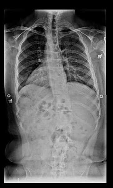
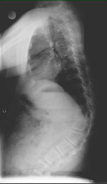
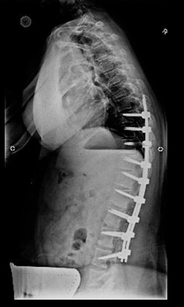
Thoracolumbar kyphosis presents in most children with achondroplasia before the age of 12 months and may reach 35 to 45 degrees before the age of 18 months.3 By age two, as children become ambulatory and gain trunk strength, kyphosis resolves in approximately 75 percent of cases3. Bracing is indicated for those children whose kyphosis does not improve by age two; the author prefers an adjustable thoracolumbar orthosis (worn full time) with a pad over the apex posteriorly.4 If the kyphosis does not improve with bracing, a trial of hyperextension casting that includes the thighs may be attempted.1 Surgical treatment is an option if the kyphosis does not improve to less than 45 degrees by the age of 8 or if neurological symptoms develop (Figure 1).
 |
 |
 |
Shortened pedicles and thickened laminae predispose patients with achondroplasia to the development of spinal stenosis2,5. In addition, decreased interpedicular distance caudally makes the narrowing especially pronounced in the lumbar spine6. Patients may present with numbness and pain in the lower extremities, incontinence of the bowel or bladder, and decreased endurance while walking, and may, in fact, stop walking altogether2. They may also place a hand on their thigh and lean forward in an attempt to relieve the symptoms of stenosis by decreasing lumbar lordosis4. MRI can correlate the level of stenosis with clinical findings.
A combination of bracing and supported sitting for the infant with achondroplasia has been suggested to prevent the progression of kyphosis, thereby lowering the risk of developing spinal stenosis7,8. If a trial of conservative treatment fails, decompression and fusion is indicated to manage the stenosis. Any surgical decompression of the spine in a skeletally immature patient with achondroplasia should be accompanied by fusion in order to prevent severe kyphosis9. In adults, decompression should be accompanied by fusion in areas of kyphosis in most patients.
Surgical Management
Because of the high rate of complications, spine surgery in the achondroplastic patient should be performed by spinal surgeons familiar with the patient population and aided by intra-operative neuromonitoring.
Posterior exposure may be complicated by prominent lordosis of the distal lumbar spine; reported angles range from 45.6 to 58.8 degrees and may exceed 90 degrees10. Exposure of L5 and S1 is further complicated by the overhanging iliac wings11. The safest way to accomplish decompression is to first thin the lamina with a burr and then to carefully dissect it off the midline4.
Dural tears are not uncommon in the achondroplastic spine and may be difficult to repair given that the dura is attenuated and the nerve roots therefore tend to bulge out of the dural sac4. Dural allograft may be needed.
Given the relatively narrow spinal canal in achondroplasia, instrumentation designed to enter the canal (e.g., hooks and wires) should be avoided whenever possible1,8. Pedicle screw instrumentation is instead preferred, with the caveat that pedicle morphology in the achondroplastic spine is unique8 (Figure 1). Given that the transverse pedicle diameter is comparable in achondroplasts and non-achondroplasts, pedicle screw diameters of 5 to 7 mm may be used5. However, the sagittal pedicle diameters of the achondroplastic spine are about 5 to 8 mm narrower and pedicle screws must be sized carefully5.
The correction of thoracolumbar kyphosis, which usually involves some shortening of the posterior column of the spine, must be done slowly and with attention to any changes in neuromonitoring. Severe focal kyphosis may be treated with either pedicle subtraction osteotomy or anterior and posterior decompression. Fusion to the sacrum in achondroplasts has recently been shown to compromise function (as measured by Short Form-36 scores) no more than more proximal fusion, though fusion to the sacrum is associated with increased post-fusion pain and increased difficulty with self-care8,12.
Outcome
Spine surgery in achondroplasia can result in specific complications, the most common of which are neurologic deficits, dural tears with cerebrospinal fluid leaks, and stenosis at other levels of the spine2. Neurologic deficits have been commonly reported in series of achondroplastic deformity correction. Cerebrospinal leaks need to be treated with secure closure with or without application of a dural patch4. The risk of stenosis at adjacent levels may be decreased by including all potential levels, usually extending past the twelfth thoracic level proximally4.
2. Diastrophic Dysplasia
Introduction
Diastrophic dysplasia (DD) is an autosomal recessive condition caused by a mutation in sulfate transporter gene DTDST. Dysfunction of DTDST results in undersulfated proteoglycans, decreasing the ability of growth plate chondrocytes to respond to local growth factors and diminishing cartilage strength and resilience13. Disorders of the spine associated with DD are cervical kyphosis and thoracolumbar scoliosis. Other features include rhizomelic dwarfism, joint contractures, club feet, and widely abducted, hypermobile “hitchhiker thumbs.” There are estimated to be between 300 to 400 patients with DD worldwide, with most believed to be residing in Finland14.
Clinical Management
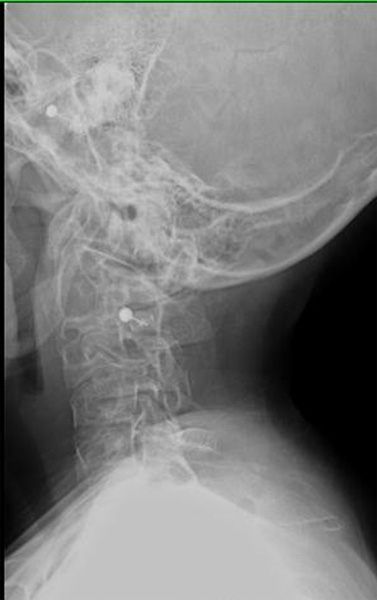
Cervical kyphosis in DD presents between infancy and the age of 6.5, averages 35 degrees (with a range of 6 to 130 degrees), and is always associated with hypoplasia of the C3-C5 vertebral bodies15. Cervical kyphosis is reported to resolve spontaneously by the age of 7, on average, if the curve is initially less than 60 degrees15 (Figure 2).
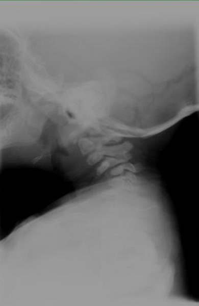 |
 |
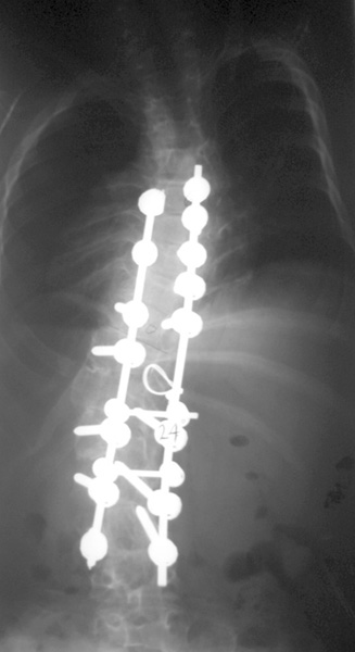
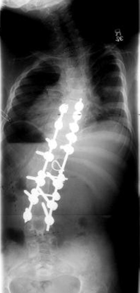
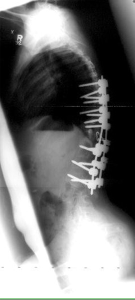
Thoracolumbar scoliosis in DD is most commonly a double major curve that is accompanied by junctional kyphosis16 (Figure 3).
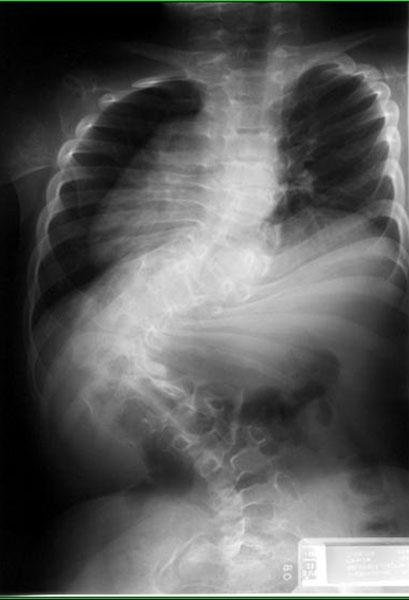 |
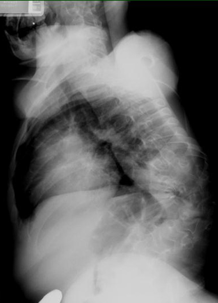 |
 |
 |
 |
Surgical Management
As long as the neurologic exam remains intact, cervical kyphosis measuring less than 60 degrees can be managed by close observation with a reasonable expectation of spontaneous resolution15.
Patients with early progressive scoliosis may present with substantial curves (greater than 60 degrees) before the age of 3 and will require surgical correction early, possibly as a staged procedure14. In idiopathic-like scoliosis, bracing of the thoracolumbar spine will not prevent the progression of curves but can be used to delay definitive fixation14.
Both posterior-only and combined anterior-posterior fusion have been described in patients with DD, with one recent series reporting significantly greater correction in patients undergoing antero-posterior surgery (40 percent correction) compared to those undergoing posterior-only surgery (13 percent correction)14,16 (Figure 3).
Outcome
Curves in DD tend to be very rigid due to premature degenerative changes in the facet joints and intervertebral discs; these curves are therefore less responsive to surgical correction16,17. Failure of fixation is seen more often in DD than in other dysplasias, most likely as a result of increased the aforementioned curve rigidity as well as the decreased size and strength of the laminae and pedicles. Pseudarthrosis has also been reported as a complication16.
Because pedicles are comparatively short, a relative spinal stenosis may result but usually does not cause symptoms17. Neurologic deficits may also be due to focal kyphosis and intrusion into the spinal canal by hooks. For this reason, pedicle screws are preferred when they can be used.
3. Spondyloepiphyseal Dysplasia
Introduction
Spondyloepiphyseal dysplasia (SED) has two forms: congenita (SEDC) and tarda (SEDT). SEDC is due to a variety of mutations in the COL2A1 gene that are thought to interfere with normal assembly of the type II collagen triple helix18. It is inherited in an autosomal dominant manner with an incidence of only 3 to 4 per million19. SEDT is X-linked, presenting at a later age than SEDC with a milder clinical picture. Orthopedic manifestations of SED include extreme short stature, kyphosis, scoliosis, coxa vara, equinovarus, and, in the case of SEDC specifically, delayed ossification19. Other conditions associated with SED include myopia and retinal detachment20.
Clinical Presentation
SED can present with hypoplasia of the odontoid or os odontoideum, resulting in atlanto-axial instability19. Fusion should be performed if there is any clinical or radiologic evidence of cord compression. The myelopathic presentation in children can include delayed motor development, spasticity, sleep apnea, and respiratory difficulty19. Thoracolumbar scoliosis is also seen in SED and can be managed with bracing in its early stages.
Surgical Management
All patients with SED should be screened for cervical spine instability prior to scoliosis correction surgery. Airway management may be challenging due to limited flexion of the neck, odontoid hypoplasia, and atlanto-axial instability21. Intra-operative motor and sensory monitoring of the spinal cord is recommended.
In the management of atlanto-axial instability, it is important to determine whether atlanto-axial instability can be reduced and whether the sagittal atlas diameter (SAD) is narrowed secondary to os odontoideum22. If the SAD is narrowed, a C1 laminectomy with occipital cervical fusion is recommended.
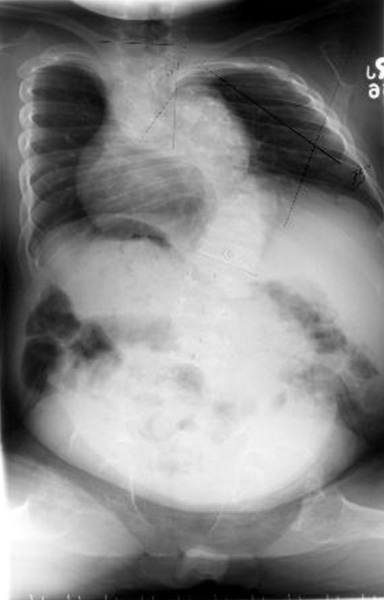
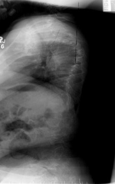
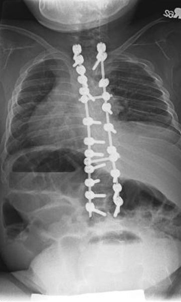
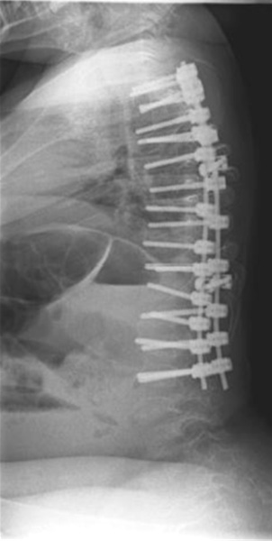
Surgery is indicated to correct thoracolumbar deformity if the curve is greater than 50 to 60 degrees, which can happen at a young age, especially in SEDC (Figure 4). The combination of a narrowed canal and small pedicles necessitates careful selection of instrumentation23. Cervical instrumentation may even be the best option for particularly diminutive vertebrae. The surgeon must strike a balance between (1) achieving adequate fixation and correction and (2) allowing for future growth. Given the limited growth potential of these small vertebrae, greater weight should be placed on the former objective4.
 |
 |
 |
 |
Outcome
Complications associated with correction of atlanto-axial instability include the inability to obtain or maintain reduction of the atlanto-axial translation, in which case decompression is needed. The arch of first cervical vertebra may be bifid; in this case, where there is an absence of adequate bone surface, fusion to the occiput may be indicated. Pre-operative CT or MRI is recommended to assess this possibility4.
II. Down Syndrome
Introduction
Down syndrome is the most common chromosome abnormality in humans, occurring in 1 of every 600 to 700 live births24. It is caused by partial or complete trisomy of chromosome 21. Disorders of the spine associated with Down syndrome include atlanto-axial and atlanto-occipital instability. Atlanto-occipital instability is attributed to flattening of the cup-shaped superior articular surface of C1 which then allows increased movement of the occipital condyles on C122. Ligamentous laxity is common in Down syndrome; laxity of the alar and transverse ligaments specifically may further contribute to instability of the upper cervical spine24. Other spine-specific conditions associated with Down syndrome include thoracolumbar scoliosis and premature spinal degeneration in early adulthood.
Clinical Presentation
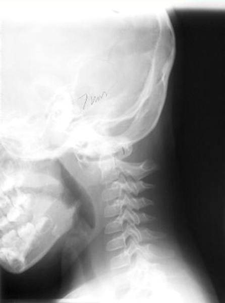
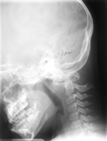
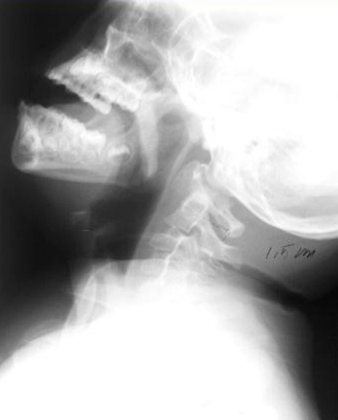
Patients with Down syndrome should be screened for cervical instability with flexion-extension radiographs (Figure 5).
 |
 |
 |
 |
Several proposals have been put forth to guide clinical management of patients based on radiographic parameters (such as the atlanto-dental interval (ADI), the distance between the posterior dens and the posterior arch of C1, and the presence of os odontoideum) but the clinical picture remains paramount25. Upper cervical instability without symptoms warrants closer monitoring of neurologic exams and restriction of activities that may place the cervical spine at risk, as well as a lower threshold for obtaining flexion-extension MRI24. Surgical intervention is recommended if neurological symptoms develop or if displacement of the atlas exceeds 8 to 10 mm.
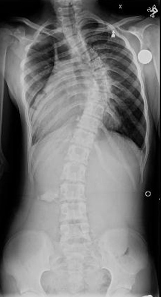
Management of scoliosis in Down syndrome is similar to management of idiopathic scoliosis; the first line of treatment is bracing followed by surgery if the curve progresses despite conservative management24 (Figure 6).
 |
 |
Surgical Management
Airway management in patients with Down syndrome may be complicated by macroglossia, mid-face hypoplasia, and instability of the cervical spine26. Up to half of patients also have cardiovascular involvement and ought to undergo cardiac evaluation prior to surgery26.
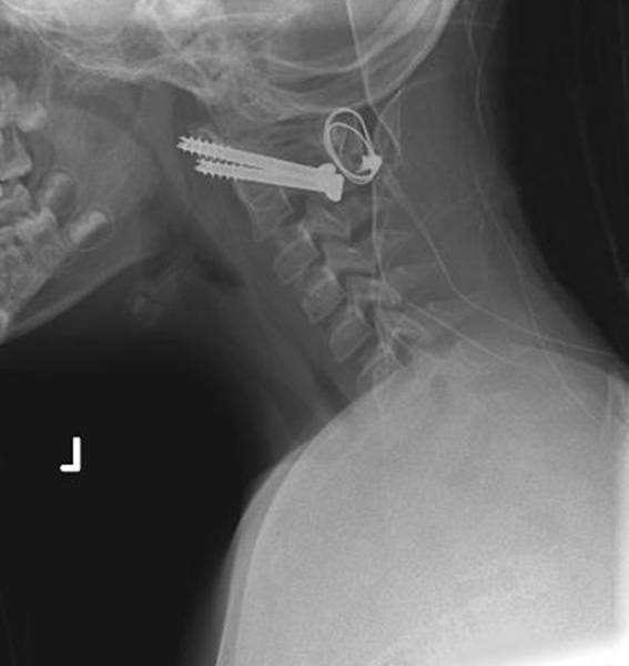
Posterior cervical fusion can be achieved via transarticular fixation of C1-C2 and occipito-cervical fusion using occipital instrumentation with bone graft. The technique for transarticular fixation of C1-C2 includes a starting point 3 to 4 mm lateral to the medial border of the C2-C3 facet27. The keys to this technique are pre-operative CT evaluation of the vertebral artery anatomy and intra-operative fluoroscopy to guide the screw towards the anterior arch of C1 at a steep trajectory27 (Figure 5). Another option is separate fixation of each level using screws in the lateral masses and pedicles24,27.
Scoliosis curves greater than 45 to 50 degrees can be managed with standard posterior instrumentation and fusion.
Outcome
Pseudarthrosis was a complication commonly associated with traditional (Gallie) fusion of the upper cervical spine. More rigid fixation has significantly decreased the rate of pseudarthrosis22,25. As alluded to above, halo fixation may help with fusion post-operatively but there is a slight risk of infection at the pin sites27. Similarly, surgical site infections are more common in Down syndrome. Other reported complications include instability in adjacent segments and neurologic deficits22.
III. Osteogenesis Imperfecta
Introduction
Osteogenesis imperfecta (OI) is caused by a variety of mutations in type I collagen genes COL1A1 and COL1A2, though several other genes have recently been implicated4,28. Genetic and clinical criteria have been used to classify OI into seven types under the expanded Sillence classification29. The most salient feature of OI is increased skeletal fragility that leads to frequent fractures, and eventually to limb deformity and disability30. Spinal disorders associated with OI include basilar invagination, kyphosis, scoliosis, spondylolysis, and spondylolisthesis.
Clinical Presentation
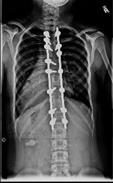
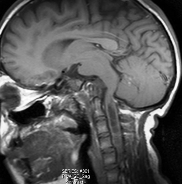
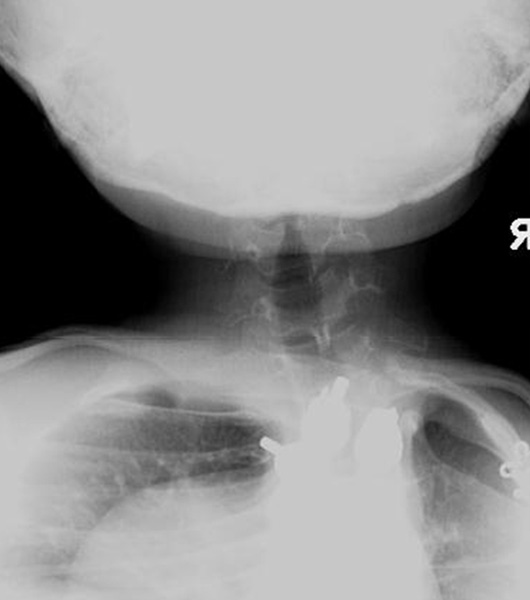
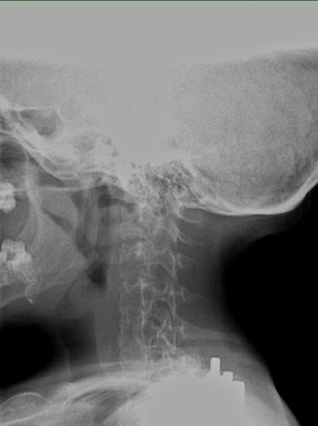
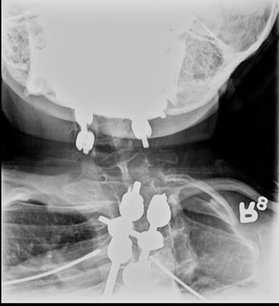
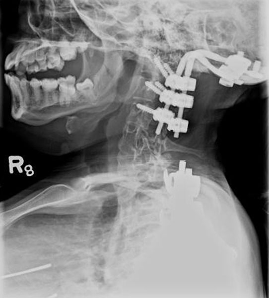
Basilar invagination occurs when the cranium settles on the upper cervical spine and the atlas and dens subsequently ascend into the foramen magnum (Figure 7). Affected patients may present with headaches, ataxia, and hypotonia, and should be followed with MRI30. Though rare, basilar invagination can lead to further neurologic dysfunction, hydrocephalus, and death when left untreated30.
 |
 |
 |
 |
 |
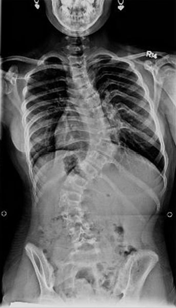
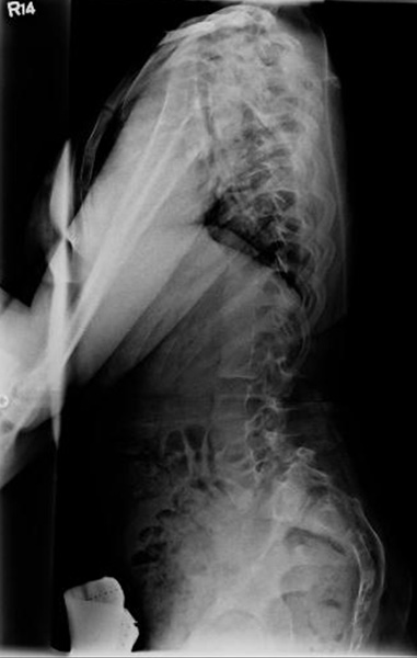
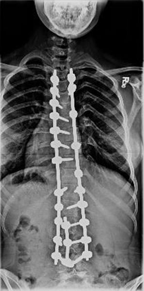
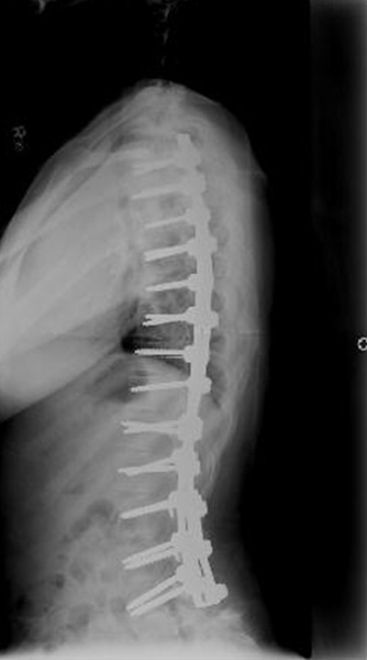
Estimates of the incidence of scoliosis in OI range from 39 to 80 percent31. Low bone mineral density (BMD) and biconcave vertebrae have been associated with an increased risk of progression of scoliosis. Conservative management with bracing may be worth a trial in young patients with type I OI and mild curves but is not often successful30. For curves greater than 50 degrees, surgery may be indicated (Figure 8) and pre-treatment with bisphosphonates for up to a year prior to surgery can increase BMD4.
 |
 |
 |
 |
The surgeon should also be aware that not all patients with OI can be helped by spinal fusion. Those with minimal or absent long-bone cortices or “crumpled” ribs may not support an implant.
Surgical Management
Pre-operative assessment of patients with OI should include lateral radiographs or MRI of the cervical spine in order to rule out basilar invagination, if the patient has not already had these studies. Basilar invagination can be managed with sub-occipital decompression (anteriorly, if necessary), upper cervical laminectomy, and occipito-cervical fusion27 (Figure 7).
Pulmonary function, and history of respiratory disease should be assessed in order to estimate the likelihood that post-operative ventilatory support will be necessary; this assessment is particularly important for patients with curves exceeding 60 degrees31.
Scoliosis correction in OI is achieved by posterior instrumentation and fusion. Surgical exposure may be complicated by a posterior flaring of the ribs that forms a “valley” by which posterior spinal elements may be obscured4. Because the pedicles are thinner or irregular in cross-section and therefore difficult to image with fluoroscopy, pedicle depths should be estimated pre-operatively using radiographs since tactile and visual feedback may be limited intra-operatively. Screws should be inserted into as many pedicles as possible to distribute the load (Figure 8). The rod chosen should have a stiffness commensurate with the expected correction of the spine; excessive rigidity increases the risk of anchor cut-out4. In certain cases, rods from cervical or pediatric applications are most appropriate. Vertebral augmentation with cement may be needed in some cases. Extensive allograft is employed in order to bring normal collagen into the region.
In rare cases where significant correction of kyphosis is indicated, anterior column support is used to prevent cut-out of the implants. The authors try to avoid post-operative bracing in these patients because it is not only mechanically ineffective but also cumbersome.
The surgeon should also be aware that dural ectasia in patients with OI renders them susceptible to dural tears and leakage of cerebrospinal fluid.
Outcomes
Some authors have advocated pre-operative halo traction placement to aid in the correction of substantial curves, reporting a success rate of 80 percent for 20 patients managed by traction prior to posterior spinal fusion32. Post-operative activity restriction or even bracing for 3 to 6 months may also be necessary to achieve the optimal outcome. Potential surgical complications include breakage of laminae or pedicles by implants, instrumentation failure post-operatively resulting in loss of correction, excessive blood loss, and late pseudarthrosis.
IV. Larsen Syndrome
Introduction
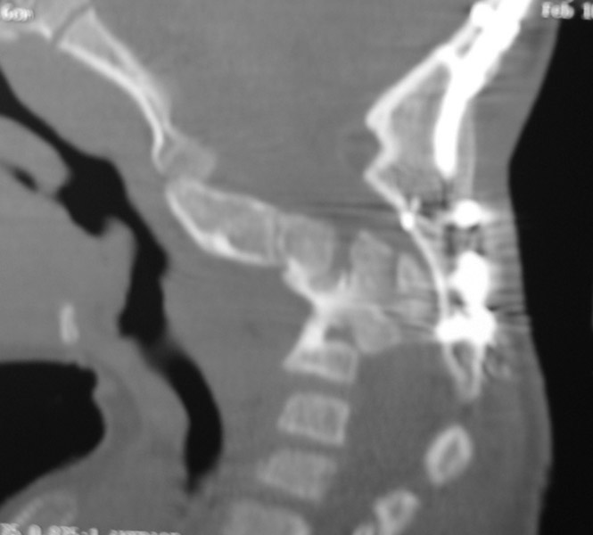
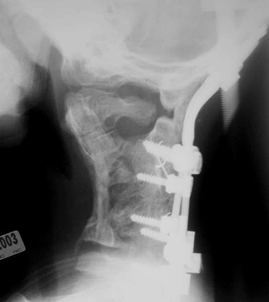
Larsen syndrome is characterized by dislocations of the knee, hip, and elbows; equinovarus or equinovalgus; and dysmorphic facies33,34. It has an estimated incidence of 1 in 100,000 births and has been associated with mutations in the filamin B (FLNB) gene on chromosome 334. Spinal deformities in Larsen syndrome include thoracolumbar scoliosis and cervical kyphosis caused by cervical spina bifida35 (Figure 9). The apex of the cervical kyphosis is usually at the fourth or fifth cervical vertebra.
 |
 |
Clinical Management
Unlike in diastrophic dysplasia, cervical kyphosis in Larsen syndrome does not improve with time and can progress to cervical dislocation and even spondyloptosis33. Initial evaluation of the child with Larsen syndrome should include radiographs and MRI of the cervical spine. As soon as deformity is identified early posterior fusion of the cervical kyphosis is indicated in infancy or early childhood in order to prevent quadriparesis33.
Surgical Management
During intubation, the anesthesiologist should try to avoid further flexion of the cervical spine in order to prevent compression from the anterior side. Other anesthetic complications that have been reported in children with Larsen syndrome include malignant hyperthermia36.
The spinal cord should be monitored during positioning and during surgery. Dissection posteriorly should be performed carefully in order to avoid cord damage at the levels with open laminae4. Localization of levels should be precise in order to avoid extension to levels above or below the desired fusion4 (Figure 9). Anteriorly, if corpectomy is indicated, it should begin at the upper and lower end of the gibbus (where the cord is least compressed) so that adequate perspective can be obtained4. If portions of the cartilage components of the vertebral bodies are left in place, they can later enlarge and lead to recurrent compression.
Outcome
To prevent the progression of deformity post-operatively, recent reports recommend posterior spinal fusion only for patients with mild or flexible kyphosis and circumferential fusion for patients with severe or rigid kyphosis greater than approximately 60 degrees33.The cervical spine should be placed in a halo vest post-operatively until adequate fusion is seen.
V. Neurofibromatosis
Introduction
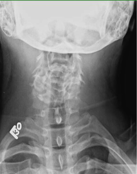
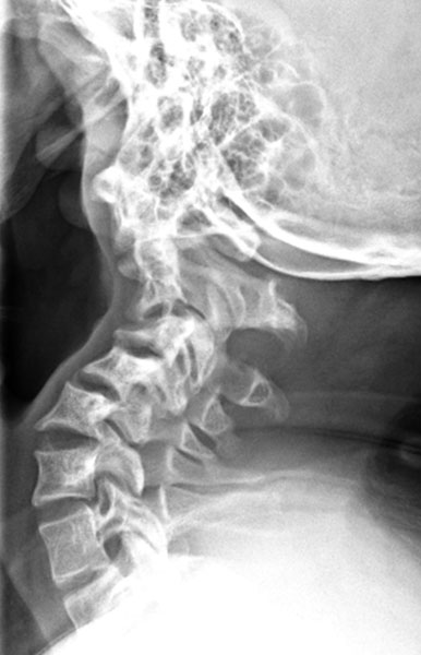
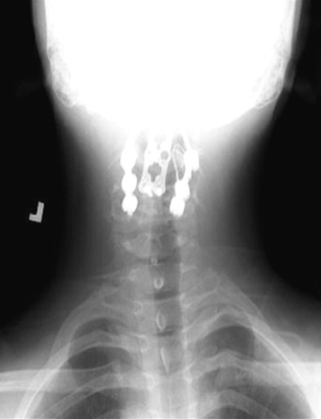
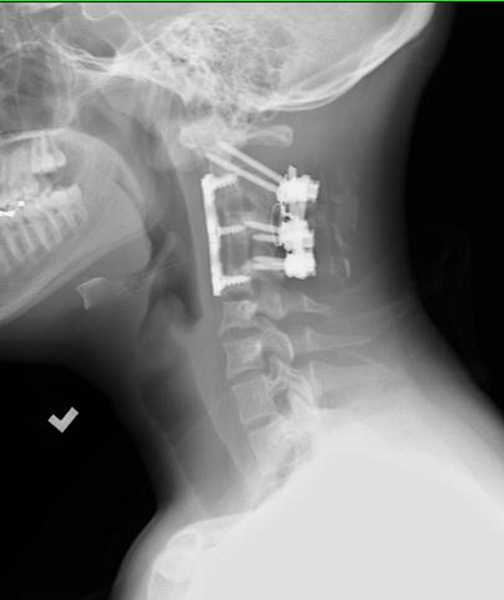
Neurofibromatosis type 1 (NF-1) is a systemic disease caused by mutation of the NF1 tumor suppressor gene on chromosome 1737. NF1 codes for neurofibromin, a mediator of proteins in the Ras pathway37. The disorder is inherited in an autosomal dominant fashion and occurs in 1 in 2,500 births37. Diagnosis of NF-1 is made clinically based on the presence of at least 2 of the diagnostic criteria established by the National Institutes of Health Consensus Development Conference: six or more café-au-lait spots greater than 5 mm before puberty or greater than 15 mm after puberty; 2 or more neurofibromas or at least 1 plexiform neurofibroma; axillary or inguinal freckling; optic glioma; 2 or more Lisch nodules; distinctive bony lesions; or an affected first-degree relative meeting the former criteria38. Spinal disorders associated with NF-1 include scoliosis, dural ectasia, and (infrequently) cervical kyphosis (Figure 10). These and other musculoskeletal complications tend to present between the ages of 5 and 10 years39.
 |
 |
 |
 |
Clinical Management
Scoliosis curves in NF-1 are characterized as either dystrophic or non-dystrophic40.
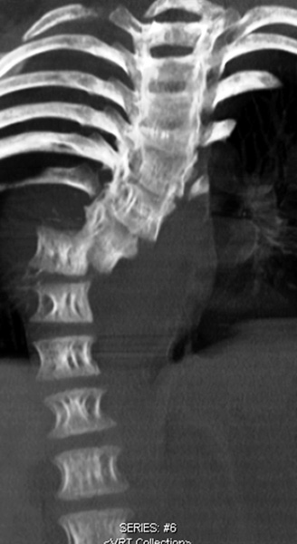
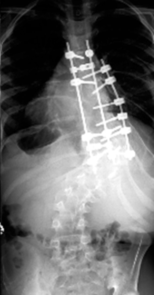
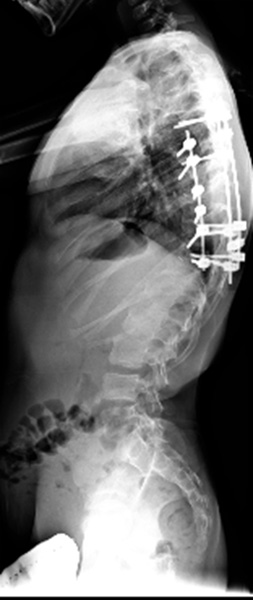
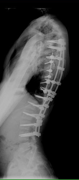
The dystrophic curve is usually sharply angulated, involving 6 or fewer vertebrae. Radiographic findings may include rib penciling, vertebral scalloping or wedging, and apical vertebral rotation39. Dystrophic scoliosis in NF-1 presents at an early age, progresses rapidly, and responds poorly to bracing39. Factors predicting increased risk of progression in dystrophic curves include a high Cobb angle, kyphosis, penciling of at least 4 ribs, and a mid- to distal thoracic apex. Patients with 3 or more dystrophic features are approximately 14 times more likely to require surgery40. Curves less than 20 degrees can be observed at 6-month intervals; surgery is indicated for curves greater than 40 degrees (Figure 11).
Non-dystrophic scoliosis in NF-1 clinically resembles idiopathic scoliosis, albeit with an earlier age of onset, a higher risk of curve progression, and a higher rate of pseudarthrosis following spinal fusion39. Non-dystrophic curves less than 25 degrees may be observed with the caveat that such curves may develop dystrophic features, especially if scoliosis presents before the age of 739,41. Curves greater than 25 degrees are amenable to bracing; however surgery is indicated for curves greater than 40 degrees39.
 |
 |
 |
|
 |
Surgical Management
A pre-operative MRI is indicated to assess the entire spinal canal for tumors, compression, dural ectasia, and intra-spinal rib dislocations4,39. Pre-operative assessment should also include radiographs of the cervical spine to prevent inadvertent cord injury under general anesthesia41. Blood loss is generally greater in NF-1 compared to the general population, especially with entry into the spinal canal, and dural ectasia increases the risk of dural tears39.
Cervical kyphosis may be managed posteriorly alone if dystrophic changes are not severe; if the kyphosis is greater than 50 degrees, anterior and posterior approaches may be employed41 (Figure 10). Pre-operative halo traction may help in severe curves if the kyphosis is not focal. In addition to a pre-operative MRI, skull radiographs should be obtained prior to the application of any form of traction in patients with NF-1 due to the risk of skull defects39.
Non-dystrophic curves greater than 40 degrees can be managed with posterior spinal fusion and instrumentation. In children younger than 9 years of age, a growth-guiding approach is usually successful. In the presence of severe dystrophic changes (including curves measuring greater than 90 degrees), both the corrective potential and the extent of fixation are limited by an exclusively posterior approach; combined surgery produces more predictable results if fixation in a dystrophic curve seems like it will be challenging41 (Figure 11). Dystrophic curves are often associated with intra-spinal neurofibromas (known as “dumbbell lesions”) which not only limit surgical exposure but also increase the risk of intra-operative bleeding39,42. Some surgeons prefer vertebral column resection. Pedicles are thinned in the dystrophic portion of the spine that threatens to complicate the placement of pedicle screws39. The fusion mass in the dystrophic portion of the curve is also thinned.
Outcome
Reported rates of pseudarthrosis range from 15 to 20 percent39,41. If a staged anterior-posterior procedure is planned, placement of a feeding tube and hyperalimentation are recommended39.
VI. Connective Tissue Disorders
1. Marfan Syndrome
Introduction
Marfan syndrome (MFS) is a systemic connective tissue disorder most frequently caused by mutations in the fibrillin-1 gene (FBN1) on chromosome 1543. Disease pathogenesis is attributed specifically to the increased levels of TGF-β associated with fibrillin-1 deficiency. Inheritance is autosomal dominant (though phenotypic expression varies widely) with an estimated prevalence of 2 to 3 per 10,000 individuals44. The diagnosis of MFS is based on the recently revised Ghent criteria that privilege two of the most salient features of the syndrome, increased aortic root diameter and ectopia lentis; the new nosology also heavily weights a known FBN1 mutation or a family history of MFS45. Other characteristic findings in the skeletal, ocular, cardiovascular, pulmonary, integumentary, and neurologic systems each receive between 1 to 3 points; a total score greater than or equal to 7 defines “systemic involvement”45. Features in the skeletal system include pectus carinatum (2 points), pectus excavatum (1 point), protusio acetabuli (2 points), and pes planus (1 point); dural ectasia merits 2 points while scoliosis and thoracolumbar kyphosis are each given 1 point45.
Clinical Management
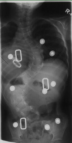
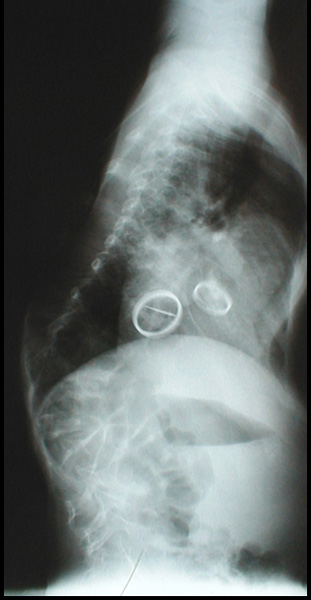
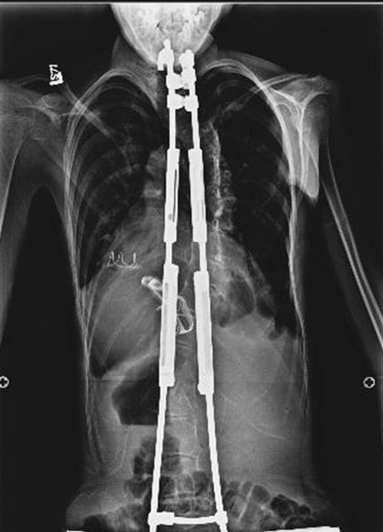
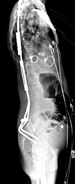
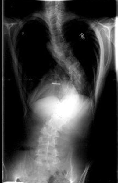
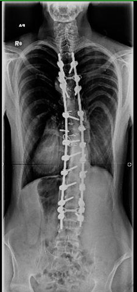
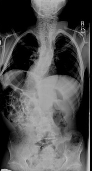
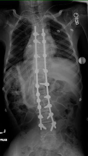
Scoliosis is present in 63 percent of patients with MFS, kyphosis in approximately 40 percent; in the sagittal plane, curves may be increased, decreased, or even reversed44. The progression of scoliosis in MFS is faster than in the general population; curves that present before the age of 3 progress most rapidly44. Unfortunately, bracing is not as effective in MFS as in idiopathic scoliosis. For curves between 15 and 40 degrees, bracing may be used in order to prevent or at least to delay the need for surgical intervention. Curves greater than 40 degrees, however, will almost certainly progress and bracing is therefore not recommended as more than a temporizing measure44. Managing infantile scoliosis in MFS is particularly challenging; bracing can be used to promote upright posture if there is concomitant kyphosis but surgical correction with growing rods will eventually become necessary46 (Figure 12).
 |
 |
 |
 |
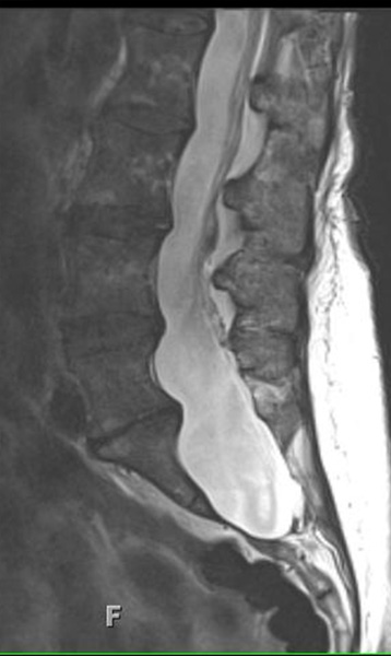
Dural ectasia—a ballooning or widening of the dural sac in the lumbosacral spine—is found in 63 to 92 percent of MFS patients47 (Figure 13). Increased hydrostatic pressure in the lumbosacral spine is hypothesized to cause expansion of already attenuated dural tissue44. Dural ectasia, in turn, contributes to the increased vertebral scalloping, reduced pedicle width, and reduced laminar thickness seen in MFS44. In adults with MFS, dural ectasias do not seem to increase in size over time; their presence is also not associated with the onset or progression of spondylolisthesis47. Spondylolisthesis is, nonetheless, approximately twice as common in MFS as in the general population (6 percent versus 3 percent) and tends to be of higher grade47.
 |
 |
 |
Surgical Management
Spine deformity surgery is required in 10 to 15 percent of individuals with MFS44. Recent evidence suggests that there is no significant difference between patients with MFS and those with adolescent idiopathic scoliosis with respect to blood loss during spinal deformity correction43. Nonetheless cardiac clearance should be obtained pre-operatively; intra-operatively, the thin antero-posterior diameter of the chest cavity of MFS patients should be kept in mind in order to avoid undue pressure on the ribs and therefore the heart4.
The presence of dural ectasia contributes to an increased rate of intra-operative cerebrospinal fluid leaks in MFS43. Pre-operative MRI or CT imaging should be obtained in order to assess the bony anatomy and to delineate any ectasia. Positioning the patient in slight Trendelenburg position may decrease the prominence of the ectasia and minimize the risk of dural tears as will minimizing dissection within the spinal canal4. If a dural tear occurs (as happens in 8 percent of spinal fixations in MFS), closure can be attempted, a dural patch placed, and bed-rest continued for two to three days post-operatively44,47.
The thin pedicles and laminae in certain regions of the spine in MFS can also make fixation challenging; pedicle screw fixation will require fluoroscopy and the use of narrow awls to find the pedicle center4. Bone mineral density, often decreased in MFS, may further undermine fixation stability.
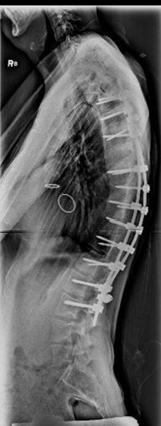
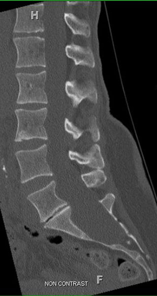
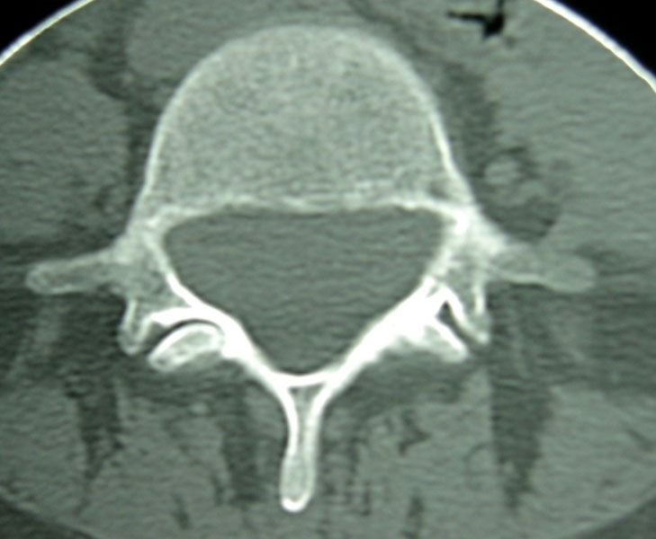
Posterior instrumentation and fusion should include all the major curves, with consideration of alignment in both sagittal and coronal planes43 (Figure 14). It is important not to fuse too short a segment of the spine; fusion to the sacrum should be considered if fusing in the lower lumbar spine, especially if distal fixation is suboptimal or if the curve is dysplastic43,44. By fusing to the sacrum, late pain and decompensation may be avoided44.
 |
 |
 |
In the patient with infantile scoliosis, growing rods are indicated if bracing fails46. Dual rods are employed, with proximal and distal fixation “foundations” robust enough to resist pullout4. A foundation of 2 vertebrae (or 3 if pedicles or laminae are dysplastic) with pairs of anchors fused proximally and distally, has proven satisfactory in our center4. If significant coronal imbalance exists or if the laminae or pedicles are not sufficient for solid fixation, the ilium is another option distally. Distraction is then performed once or twice per year4. The initial distraction followed by 5 years of growing treatment can reasonably be expected to produce at least 11 cm of additional spine length46.
Outcome
Rates of hook dislodgment, pseudarthrosis, and fixation failure in MFS have been reported to be 22 percent, 10 percent, and 21 percent, respectively44. The failure of fixation may be due to attenuated pedicles and laminae, decreased bone mineral density, and ligamentous laxity. For these reasons, the revision rate is significantly higher than in AIS43.
Because valvular incompetence and cardiac failure have been reported in the post-operative period, patients with MFS should also be followed by the cardiologist or a medical team4. In addition, superior mesenteric artery syndrome has been associated with the correction of significant kyphosis in MFS4. Other post-operative considerations include pneumothorax (a spontaneous occurrence in MFS) and hemothorax.
2. Ehlers-Danlos Syndrome
Introduction
Ehlers-Danlos syndrome (EDS) is a connective tissue disorder caused by defects in the synthesis and post-translational modification of fibrillar collagen types I, III, and V48. The Berlin system identified nine types of EDS. The more recent Villefranche classification system describes six subtypes of EDS, grouped according to clinical, genetic, and biochemical features48. The incidence of EDS is estimated to be from 1 in 25,000 to 1 in 5,000 births. The most prominent features of EDS are hyperelastic skin, hyperextensible joints, and easy bruising (due to vascular fragility)49. Scoliosis is most common in types I (classic) and VI (ocular-scoliotic). However, spinal deformity does not appear to be as common as in Marfan syndrome4. Spondylolisthesis can also be seen in EDS.
Clinical Management
The scoliosis seen in EDS can become rigid and severe49. The role and outcomes of bracing in EDS patients have not been well studied but the few mentions made in the literature suggest bracing does little to slow disease progression50,51. Surgical correction is offered for curves greater than 50 to 60 degrees4. Appropriate assessment of vascular structures in type IV EDS with MRI and angiography can aid pre-operative planning49.
Surgical Management
Pre-operative MRI can also be used to evaluate patients for dural ectasia, the presence of which increases the risk of dural tears in the lumbosacral spine4.
The anesthesiologist should be aware of the increased blood loss caused by vascular fragility in EDS; reports of mean estimated blood loss range from 1,243 mL to 2,800 mL, depending on the surgical approach51. The posterior approach is associated with blood loss at the lower end of this range (mostly from small muscular and periosteal vessels) and is therefore preferred for deformity correction in EDS4. Because of the increased risk of significant vascular injury (e.g., to iliac vessels and segmental branches of the aorta), anterior release should be avoided whenever possible49. If anterior vessel injury is suspected, the prudent approach is to stage the procedure, with the assistance of a vascular surgery consult. Staged, temporary internal distraction followed by segmental instrumentation may decrease rigidity and avoid the need for an anterior procedure49.
Neurologic deficits during spinal deformity correction may be more common in EDS; proposed mechanisms for this phenomenon include increased ligamentous laxity that allows for greater spinal cord distraction and increased vascular fragility that compromises perfusion of the cord4.
Outcome
Adequate deformity correction can be obtained in EDS but may be complicated by junctional kyphosis49. Post-operative wound hematoma is common and can be minimized by the placement of a wound drain4.
VII. Stickler Syndrome
Introduction
At least four different varieties of Stickler syndrome (or hereditary arthro-ophthalmopathy) have been described. The membranous (type 1 vitreous) form of Stickler syndrome is associated with mutations in the COL2A1 gene while the beaded vitreous (type 2) and non-ocular (type 3) forms are associated with mutations in COL11A1 and COL11A2, respectively52. Types 1, 2, and 3 all share autosomal dominant patterns of inheritance, and a recessive form caused by a mutation in the COL9A1 gene has been recently described53. The total incidence of all forms of Stickler syndrome is approximately 1 in 8,000, though it is suggested that the syndrome continues to be underdiagnosed52,54.
A panel of major and minor criteria is used to diagnose Stickler syndrome55. Characteristic craniofacial abnormalities include cleft palate or bifid uvula, micrognathia, and mid-face hypoplasia56. Ocular defects, including myopia and retinal detachment, are found in all forms except type 3 (because COL11A2 is not expressed in tissues of the eye)52. Approximately 80 percent of patients with Stickler syndrome have musculoskeletal abnormalities including muscle hypoplasia, articular hypermobility, early-onset osteoarthritis, protusio acetabuli, coxa valga, and spinal anomalies55.
Clinical Manifestation
Spinal abnormalities include spondylolisthesis, scoliosis, and hyperkyphosis. In one series of 53 patients with Stickler syndrome, 18 (34 percent) were found to have scoliosis55. Of these 18 patients, 4 had curves with a left thoracic apex orientation; early MRI is indicated in the management of these cases. In general, scoliosis in Stickler syndrome can be managed conservatively with bracing; surgery is indicated if the curve does not correct (Figure 15). In that same study, only 1 of the 18 patients with scoliosis underwent surgical correction55.
 |
 |
Focal thoracic kyphosis was also found in 43 percent of these patients55. Endplate abnormalities such as Schmorl’s nodes and platyspondylia are also common in Stickler syndrome55. Back pain affects up to 85 percent of adult patients, with a younger age of presentation compared to the general population55.
Surgical Management
Recent evidence suggests that valvular abnormalities are no more prevalent in patients with Stickler syndrome than in the general population, as was once thought, and that routine pre-operative echocardiography and antibiotic prophylaxis are therefore no longer indicated57. The anesthesiologist should, however, be prepared to manage an airway potentially complicated by micrognathia or mid-face hypoplasia4. Standard posterior instrumentation and fusion may be used in the correction of spinal deformity.
Outcome
To date, case series on outcomes of spinal deformity correction in Stickler syndrome have not been published. There is an isolated report of successful correction of thoracolumbar scoliosis with posterior spinal fusion and instrumentation55.
References
1. Shirley ED, Ain MC. Achondroplasia: manifestations and treatment. J Am Acad Orthop Surg. 2009;17(4):231–241.
2. Ain MC, Chang T-L, Schkrohowsky JG, Carlisle ES, Hodor M, Rigamonti D. Rates of perioperative complications associated with laminectomies in patients with achondroplasia. J Bone Joint Surg Am. 2008;90(2):295–298.
3. Borkhuu B, Nagaraju DK, Chan G, Holmes L Jr, Mackenzie WG. Factors related to progression of thoracolumbar kyphosis in children with achondroplasia: a retrospective cohort study of forty-eight children treated in a comprehensive orthopaedic center. Spine. 2009;34(16):1699–1705.
4. Sponseller PD, Mesfin A. Spine deformity secondary to dysplasias and other diseases. In: Kim DH, ed. Surgery of the pediatric spine. 1st ed. New York: Thieme; 2008:896.
5. Srikumaran U, Woodard EJ, Leet AI, Rigamonti D, Sponseller PD, Ain MC. Pedicle and spinal canal parameters of the lower thoracic and lumbar vertebrae in the achondroplast population. Spine. 2007;32(22):2423–2431.
6. Vleggeert-Lankamp C, Peul W. Surgical decompression of thoracic spinal stenosis in achondroplasia: indication and outcome: Clinical article. J Neurosurg Spine. 2012;17(2):164–172.
7. Schkrohowsky JG, Hoernschemeyer DG, Carson BS, Ain MC. Early presentation of spinal stenosis in achondroplasia. J Pediatr Orthop. 2007;27(2):119–122.
8. Shirley ED, Ain MC. Kyphotic Deformities in Skeletal Dysplasias. Semin Spine Surg. 2012;24(3):186–191.
9. Baca KE, Abdullah MA, Ting BL, et al. Surgical decompression for lumbar stenosis in pediatric achondroplasia. J Pediatr Orthop. 2010;30(5):449–454.
10. Karikari IO, Mehta AI, Solakoglu C, Bagley CA, Ain MC, Gottfried ON. Sagittal spinopelvic parameters in children with achondroplasia: identification of 2 distinct groups: Clinical article. J Neurosurg Spine. 2012;17(1):57–60.
11. Uematsu S, Wang H, Kopits SE, Hurko O. Total craniospinal decompression in achondroplastic stenosis. Neurosurgery. 1994;35(2):250–257; discussion 257–258.
12. Farmer KW, Brinkley MF, Skolasky RL, Sponseller PD, Ain MC. Lumbar fusion in achondroplasia: does fusion to the sacrum affect function? J Pediatr Orthop. 2009;29(5):476–480.
13. Weiner DS, Jonah D, Kopits S. The 3-dimensional configuration of the typical hip and knee in diastrophic dysplasia. J Pediatr Orthop. 2010;30(4):403–410.
14. Jalanko T, Remes V, Peltonen J, Poussa M, Helenius I. Treatment of spinal deformities in patients with diastrophic dysplasia: a long-term, population based, retrospective outcome study. Spine. 2009;34(20):2151–2157.
15. Remes V, Marttinen E, Poussa M, Kaitila I, Peltonen J. Cervical kyphosis in diastrophic dysplasia. Spine. 1999;24(19):1990–1995.
16. Matsuyama Y, Winter RB, Lonstein JE. The spine in diastrophic dysplasia. The surgical arthrodesis of thoracic and lumbar deformities in 21 patients. Spine. 1999;24(22):2325–2331.
17. Remes V, Tervahartiala P, Poussa M, Peltonen J. Thoracic and lumbar spine in diastrophic dysplasia: a clinical and magnetic resonance imaging analysis. Spine. 2001;26(2):187–195.
18. Online Mendelian Inheritance in Man, OMIM. Johns Hopkins University, Baltimore, MD. MIM Number: 183900: September 24, 2012. 2012.
19. Song D, Maher CO. Spinal disorders associated with skeletal dysplasias and syndromes. Neurosurg Clin N Am. 2007;18(3):499–514.
20. Veeravagu A, Lad SP, Camara-Quintana JQ, Jiang B, Shuer L. Neurosurgical Interventions for Spondyloepiphyseal Dysplasia Congenita: Clinical Presentation and Assessment of the Literature. World Neurosurg. 2012.
21. Lin C-P, Su C-F, Lin W-Y, et al. Modified lightwand intubation in a child with spondyloepiphyseal dysplasia congenita. Acta Anaesthesiol Taiwanica Off J Taiwan Soc Anesth. 2011;49(2):66–68.
22. Miyoshi K, Nakamura K, Haga N, Mikami Y. Surgical treatment for atlantoaxial subluxation with myelopathy in spondyloepiphyseal dysplasia congenita. Spine. 2004;29(21):E488–491.
23. Schwend RM, Nandyala SV. Current State of Pedicle Screw Constructs in Children with Spinal Deformity. Semin Spine Surg. 2012;24(3):192–201.
24. Caird MS, Wills BPD, Dormans JP. Down syndrome in children: the role of the orthopaedic surgeon. J Am Acad Orthop Surg. 2006;14(11):610–619
25. Hwang SW, Jea A. A review of the neurological and neurosurgical implications of down syndrome in children. Clin Pediatr (Phila). 2013;52(9):845–856.
26. Farag E. Anesthesia for Spine Surgery. Cambridge University Press; 2012.
27. Reilly CW, Choit RL. Transarticular screws in the management of C1-C2 instability in children. J Pediatr Orthop. 2006;26(5):582–588.
28. Grover M, Campeau PM, Lietman CD, et al. Osteogenesis imperfecta without features of type V caused by a mutation in the IFITM5 gene. J Bone Miner Res Off J Am Soc Bone Miner Res. 2013.
29. Rauch F, Glorieux FH. Osteogenesis imperfecta. Lancet. 2004;363(9418):1377–1385.
30. Forlino A, Cabral WA, Barnes AM, Marini JC. New Perspectives on Osteogenesis Imperfecta. Nat Rev Endocrinol. 2011;7(9):540–557.
31. Widmann RF, Bitan FD, Laplaza FJ, Burke SW, DiMaio MF, Schneider R. Spinal deformity, pulmonary compromise, and quality of life in osteogenesis imperfecta. Spine. 1999;24(16):1673–1678.
32. Janus GJ, Finidori G, Engelbert RH, Pouliquen M, Pruijs JE. Operative treatment of severe scoliosis in osteogenesis imperfecta: results of 20 patients after halo traction and posterior spondylodesis with instrumentation. Eur Spine J Off Publ Eur Spine Soc Eur Spinal Deform Soc Eur Sect Cerv Spine Res Soc. 2000;9(6):486–491.
33. Sakaura H, Matsuoka T, Iwasaki M, Yonenobu K, Yoshikawa H. Surgical treatment of cervical kyphosis in Larsen syndrome: report of 3 cases and review of the literature. Spine. 2007;32(1):E39–44.
34. Zhang D, Herring JA, Swaney SS, et al. Mutations responsible for Larsen syndrome cluster in the FLNB protein. J Med Genet. 2006;43(5):e24.
35. De la Rocha A, Birch JG, Schiller JR. Precocious appearance of the capital femoral ossific nucleus in Larsen syndrome. J Bone Joint Surg Am. 2012;94(9):e55.
36. Ghaffaripour S, Ghahramaninejad F, Shahbazi S. Malignant hyperthermia in Larsen syndrome. Paediatr Anaesth. 2009;19(9):927–928.
37. Pasmant E, Vidaud M, Vidaud D, Wolkenstein P. Neurofibromatosis type 1: from genotype to phenotype. J Med Genet. 2012;49(8):483–489.
38. Neurofibromatosis. Conference statement. National Institutes of Health Consensus Development Conference. Arch Neurol. 1988;45(5):575–578.
39. Crawford AH, Parikh S, Schorry EK, Von Stein D. The immature spine in type-1 neurofibromatosis. J Bone Joint Surg Am. 2007;89 Suppl 1:123–142.
40. Lykissas MG, Schorry EK, Crawford AH, Gaines S, Rieley M, Jain VV. Does the presence of dystrophic features in patients with type 1 neurofibromatosis and spinal deformities increase the risk of surgery? Spine. 2013;38(18):1595–1601.
41. Delucia TA, Yohay K, Widmann RF. Orthopaedic aspects of neurofibromatosis: update. Curr Opin Pediatr. 2011;23(1):46–52.
42. Feldman DS, Jordan C, Fonseca L. Orthopaedic manifestations of neurofibromatosis type 1. J Am Acad Orthop Surg. 2010;18(6):346–357.
43. Gjolaj JP, Sponseller PD, Shah SA, et al. Spinal deformity correction in Marfan syndrome versus adolescent idiopathic scoliosis: learning from the differences. Spine. 2012;37(18):1558–1565.
44. Demetracopoulos CA, Sponseller PD. Spinal deformities in Marfan syndrome. Orthop Clin North Am. 2007;38(4):563–572, vii.
45. Loeys BL, Dietz HC, Braverman AC, et al. The revised Ghent nosology for the Marfan syndrome. J Med Genet. 2010;47(7):476–485.
46. Sponseller PD, Thompson GH, Akbarnia BA, et al. Growing rods for infantile scoliosis in Marfan syndrome. Spine. 2009;34(16):1711–1715.
47. Mesfin A, Ahn NU, Carrino JA, Sponseller PD. Ten-year clinical and imaging follow-up of dural ectasia in adults with Marfan syndrome. Spine J Off J North Am Spine Soc. 2013;13(1):62–67.
48. Callewaert B, Malfait F, Loeys B, De Paepe A. Ehlers-Danlos syndromes and Marfan syndrome. Best Pract Res Clin Rheumatol. 2008;22(1):165–189.
49. Yang JS, Sponseller PD, Yazici M, Johnston CE 2nd. Vascular complications from anterior spine surgery in three patients with Ehlers-Danlos syndrome. Spine. 2009;34(4):E153–157.
50. McMaster MJ. Spinal deformity in Ehlers-Danlos syndrome. Five patients treated by spinal fusion. J Bone Joint Surg Br. 1994;76(5):773–777.
51. Rabenhorst BM, Garg S, Herring JA. Posterior spinal fusion in patients with Ehlers-Danlos syndrome: a report of six cases. J Child Orthop. 2012;6(2):131–136.
52. Baker S, Booth C, Fillman C, et al. A loss of function mutation in the COL9A2 gene causes autosomal recessive Stickler syndrome. Am J Med Genet A. 2011;155A(7):1668–1672.
53. Hoornaert KP, Vereecke I, Dewinter C, et al. Stickler syndrome caused by COL2A1 mutations: genotype-phenotype correlation in a series of 100 patients. Eur J Hum Genet EJHG. 2010;18(8):872–880.
54. Rose PS, Levy HP, Liberfarb RM, et al. Stickler syndrome: clinical characteristics and diagnostic criteria. Am J Med Genet A. 2005;138A(3):199–207.
55. Rose PS, Ahn NU, Levy HP, et al. Thoracolumbar spinal abnormalities in Stickler syndrome. Spine. 2001;26(4):403–409.
56. Richards AJ, McNinch A, Martin H, et al. Stickler syndrome and the vitreous phenotype: mutations in COL2A1 and COL11A1. Hum Mutat. 2010;31(6):E1461–1471.
57. Ahmad N, Richards AJ, Murfett HC, et al. Prevalence of mitral valve prolapse in Stickler syndrome. Am J Med Genet A. 2003;116A(3):234–237.
Authors: Crawford, Alvin H.; Jain, Viral V.
Introduction
Neurofibromatosis is a spectrum of multifaceted diseases involving not only neuroectoderm, mesoderm and endoderm. They present a wide range of clinical manifestations which may include schwannomas, neurofibromas, and/or café au lait spots. The primary pathology is believed to be a hamartomatous disorder of neural crest derivation. Common findings in pediatric NF-1 are noted on Table 11. This chapter deals with the primary and secondary effects of NF-1 on the pediatric spine.
| Manifestation | Percent |
|---|---|
| Café au lait and cutaneous tumors by adolescence | 72-90 |
| Positive family history | 45-48 |
| Central nervous system lesions | 16-26 |
| Scoliosis | 20-26 |
| Skull and facial deformities | 20 |
| Failure to thrive in infancy | 16 |
| Aqueducatal stenosis | 16 |
| Breast enlargement | 13 |
| Seizures | 13 |
| Hemihypertrophy | 13 |
| Tibial dysplasia | 12 |
| Extracranial malignancy | 12-20 |
| Vascular disease | 10 |
| Mediastinal tumors | 8 |
Diagnostic Types
Most investigators now accept four clinical forms of Neurofibromatosis: The most common form is Neurofibromatosis Type –1 (NF1) previously known as von Recklinghausen disease, which involves predominantly peripheral nervous system although it involves cells of mesodermal origin as well. It is an autosomal dominant disorder affecting about one in 4,000 persons2; multiple hyperpigmented areas (café au lait macules) and neurofibromas are characteristic. Neurofibromatosis Type-2 (NF2) is the second most common form of the disorder involving central nervous system and is associated with bilateral vestibular schwannomas and multiple spinal schwannomas. Segmental neurofibromatosis is considered to be a mosaic form of NF1 with features of NF1 present in single body segment.3 Schwannomatosis is the most recently described form manifesting itself as multiple deep painful schwannomas and is thought to represent a mosaic form of NF2.4
The 1987 Consensus Development Conference of the National Institutes of Health on NF-1 concluded that the diagnosis of von Recklinghausen's NF-1 was established when two or more of the diagnostic criteria listed in Table 2 were found5.
Other disorders of pigmentation such as McCune-Albright syndrome, LEOPARD syndrome, Noonan's syndrome and Watson's syndrome can be confused with von Recklinghausen's NF-16.
Since the consensus panel meeting, specific kinds of learning disabilities and MRI abnormalities (especially in children) have been associated specifically with NF-1. These are useful even during early life.7 More recently, anterolateral bowing of the tibia has been considered as one of the criterion for the diagnosis of NF1.8
| (1) six or more café au lait macules greater than 5 mm in widest diameter in prepubertal children and more than 15 mm in widest diameter in postpubertal individuals |
| (2) two or more neurofibromas of any type or one plexiform neurofibroma |
| (3) freckling in the axillary or inguinal regions |
| (4) optic glioma |
| (5) two or more Lisch nodules (iris hamartomas) |
| (6) a distinctive osseous lesion such as sphenoid dysplasia or thinning of a long bone cortex with or without pseudarthrosis |
| (7) a first-degree relative (parent, sibling, or offspring) with von Recklinghausen's disease identified by the above criteria. |
Molecular Genetics
NF-1 is the most common human single-gene disorder. It affects at least one million people throughout the world. It is seen in all racial and ethnic groups. It would be conservative to estimate that half of them will suffer serious medical and social complications. The gene is fairly large sized, in the range of 300,000 base pairs. (Most genes are several tens of thousands of base pairs, and the largest known, the gene for Duchenne muscular dystrophy, extends over 2.5 million base pairs.)
In 1990, the gene locus of NF-1 in humans was discovered on the long arm of chromosome 17, and its protein product, neurofibromin, was identified 9, 10. Expression of neurofibromin is highest in neurons, oligodendrocytes, non-myelinated Schwann cells, adrenal medulla, leukocytes, and testes. Neurofirbomin act as a supressor for p21-Ras, an oncogene, and therefore, the loss of neurofibromin, as in NF-1, would lead to inability to shut off activated p21-Ras, with subsequent aberrant growth promoting signals. In this respect, NF-1 gene is a typical tumor suppressor gene and NF-1 is the most common human cancer predisposition syndrome (*1,2,3)
Molecular Diagnosis
Direct sequencing of the NF1 gene is now available for genetic testing. In contrast to the previously used protein truncation assay which only detected 60-70% of NF1 mutations it detects the mutation in 95% and is considered as the gold standard.11, 12 Animal models such as knockout mouse are now available that will greatly increase our understandings of the disease and help in development of therapeutic strategies.
Clinical Findings
Café au Lait Spots
Café au lait spots are present in well over 90% of all patients with NF-1. The pigmentation is tan, macular, and melanotic in origin and is located in and around the basal layer of the epidermis; the lesions may vary in shape, size, number, and location even in areas of the skin not exposed to the sun (Fig 1D).
Axillary and Inguinal Freckling
Freckles-diffuse,small, hyperpigmented spots up to 2-3mm in diameter found in the axillary and inguinal region (areas not usually exposed to sunlight) are helpful diagnostic criteria for NF-1.
Lisch Nodules
Lisch nodules, or iris-pigmented hamartomas, are present in 94% of patients with NF-1 who are 6 years of age or older; they are not seen in non-NF-1 individuals normal.
Optic Glioma
Although optic gliomas account for only 2% to 5% of all brain tumors in childhood, as many as 70% of the cases are found in persons with NF-1. They occur in 10% of children with NF-1. Gliomas involving the optic chiasma are most likely to cause visual impairments and precocious puberty.
Neurofibromas
Neurofibromas mostly involve the skin, but they may be seen in deeper peripheral nerves. They may be nodular and discrete or diffuse with interdigitation with surrounding tissues. Highly vascular plexiform neurofibromas may cause segmental or localized hypertrophy. Puberty or pregnancy may cause an increase in the size and number of the lesions 13.
Cutaneous Neurofibroma
Cutaneous neurofibromas, formerly called fibroma molluscum, are present in subcutaneous tissues, are found only after puberty. They have been recently found to have axons and Schwann cells and now appropriately called dermal neurofibromas. (30).
Plexiform Neurofibroma
Plexiform neurofibromas are subcutaneous neurofibromas tat arise from large nerve roots and interdigitate with normal tissues. that have a ropy, “bag of worms” feeling. Their cutaneous involvement may cause decreased sensation. Underlying plexiform neurofibromas are often covered by an area of hyperpigmented skin and/or a hairy patch. They have the potential for malignant degeneration6.
Pigmented Nevi
Eight percent of all patients presenting with NF-1 have CAL spots with geographic descriptions, e.g., “nevus lateralus” and “bathing trunk” Nevi or hyperpigmentation may be present in up to 6% of children with NF-16. Some are quite sensitive and may overlay an underlying subcutaneous plexiform neurofibroma.
Elephantiasis
Frequently, large soft-tissue masses are seen in NF-1. These masses have been termed pachydermatocele or elephantiasis neuromatosa and are characterized by a rough, raised, villous type of skin hypertrophy presenting an unmistakable appearance.
Verrucous Hyperplasia
Verrucous hyperplasia is an infrequent unsightly tremendous overgrowth of the skin, with thickening of a velvety-soft papillary quality. Many crevices form and tend to break down easily, with some weeping occurring in the skin folds.
Spinal Deformities
Spinal deformities are the most common orthopedic manifestation of NF1. True incidence of spinal deformities in NF1 is not known. It is quoted as from 2% to 36% in the literature.14, 15In the NF clinic at our institution, it is 23%.2 Functional scoliosis resulting fromlimb hypertrophy or long-bone dysplasia leading to limb length inequality must be ruled out inpatients with NF1. The spinal deformities tend to develop early in the life, therefore, all pre-adolescent children with NF1 shouldbe evaluated by scoliosis screening or the Adam forward-bendtest to rule out the presence of a spinal deformity.
Traditionally, two forms of spinal deformities are described in NF1: Dystrophic and Non-Dystrophic. The distinctions between these two varieties of spinal deformities are clearer in the thoracic and thoracolumbar regions. Non-dystrophic curves are similar to idiopathic curves with some exceptions. The dystrophic changes in NF1 are described in Table 3. The cause of dystrophic changes may be intrinsic or associated with anomaliesof the spinal canal secondary to abnormalities of the spinal cord/dura matersuch as tumors, meningoceles, and/or dural ectasia.Dystrophic changes may also occur even if the intraspinal contents are normal. Several investigatorshave suggested that there is no standard pattern of spinal deformityin NF1 and that the types of curvature are variable.16, 17
| Feature | Percent |
|---|---|
| Rib penciling | 62 |
| Vertebral rotation | 51 |
| Vertebral scalloping Posterior Lateral | 31 |
| Vetebral Wedging | 36 |
| Spindling of transverse process | 31 |
| Widened inter-pedicular distance | 29 |
| Enlarged intervetebral foraminae | 25 |
Radiology
Routine posteroanterior and lateral cervical, thoracolumbar, and sacral radiographs are necessary when studying the deformed spine because of the potential for occult deformity in each location. Careful attention is directed on plain x-rays to the sagittal plane for evidence of abnormal lordosis, kyphosis or dystrophic changes.
Computed tomography (CT) has been used extensively to identify the occasional abnormal structure of the spine in NF-1; it allows one to assess the spinal canal, its contents, and the associated anatomy. Three-dimensional reconstruction enhances the explicitness of the bony detail. The addition of contrast myelography to CT has allowed the surgeon to identify lesions found in and about the spinal canal. Magnetic resonance imaging (MRI) is utilized to determine the internal contents of the spinal canal and will show the presence of lesions within and about the spinal cord itself.18
Management Of Spinal Deformities
Cervical Deformities
The cervical spine deformity is usually kyphosis (Figure 1). In our experience, the most common cause of cervical deformity is decompression laminectomies without fusion, that leads to instability with progressive kyphotic deformities. Until recently, only casual references to the cervical spine have been evident in studies of other manifestations of NF-1. Many patients with cervical spine deformities are asymptomatic and clinically not apparent. Even so curves in this area are more frequently associated with dysplastic lesions than in other areas of the spine 19. The clinical consequences of cervical NF-1 tend to be less marked than in other regions because the cord versus canal diameter is commonly reported to be less critical. Because of its generally asymptomatic nature the problems is probably under reported. However, these lesions should not be disregarded, because the tendency of the disease to progress has led to severe neurological deficits in several cases.
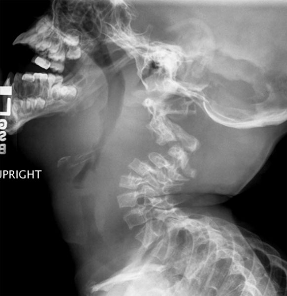 |
|
Anteroposterior and lateral radiographs of the cervical spine are recommended at the time of original evaluation of all spinal deformities in NF-1 patients. If dystrophic changes are noted, oblique radiographs to rule out dumbbell lesions (enlargement of neuroforamina due to intraspinal meningoceles, solitary tumors, or interstitial hypertrophic neuritis) and lateral flexion and extension radiographs to rule out instability should be obtained. One should be suspicious of scalloping of the vertebral bodies or an increase in the size of the neural foramina. If found, an MRI should be performed to evaluate the intraspinal contents.
Posterior spinal fusion is recommended for cervical spine deformity with instability. Because of the occasional extensive laminectomy required to completely excise the tumor and the resulting instability, posterior and anterior spinal fusion should always be carried out. If the surrounding tumor load prevents anterior exposure, an occipito-cervical fusion and stabilization may be required. Often halo vest or cast is used to assist in post-operative stabilization. Multiple view radiographs of the cervical spine are mandatory before general anesthesia or halo traction to rule out dystrophic deformity and possible skull defects.
Thoracic and Lumbar spinal deformities
Thoracic scoliosis is the most common osseous defect associated with NF 1. The cause of spinal deformity is unknown, but it has been suggested to be secondary to osteomalacia, localized neurofibromatous tumor eroding and infiltrating bone, endocrine disturbances, or mesodermal dysplasia. Two types of deformity tend to occur, dystrophic and non-dystrophic.
Non dystrophic scoliosis
This is the common variety of spinal deformity observed in NF1. These curves behave similar to idiopathic curves with some differences.20-22 This form usually involves 8 to 10 spinal segments. Most often, the deformity is convex to the right. However, these curves usually present earlier than the idiopathic curves and are more prone to progression. Furthermore, the rate of pseudoarthrosis following a fusion surgery is higher in these patients.23
The authors' recommendation for the treatment of non-dystrophic curvatures is to treat them as idiopathic scoliosis. The curve of less than 25° should be observed. Curves between 25° and 40° can be treated with braceing.24 Once beyond 40°, surgery by posterior spinal fusion is usually indicated4. Curves greater than 60° are treated with anterior release with bone-grafting, followed by an instrumented posterior spinal fusion.23 This is necessary because the curve is usually more rigid than is a similar-sized curve in idiopathic scoliosis. We recommend postoperative orthotic immobilization, although others have managed these patients without postoperative immobilization, with good early results.17
There is the distinct possibility of modulation across a spectrum of nondystrophic to dystrophic curvatures, but this tendency shows no consistent pattern 25.
Dystrophic scoliosis
This is an uncommon but malignant form of spinal deformity. It is characterized by early-onset, rapidly progression and is more difficult to treat.26, 27 Typically, the dystrophic curve is a short-segmented, sharply angulated type that includes fewer than six spinal segments. Dystrophic curves may be associated with kyphosis and have a higher incidence of neurologic injury.27, 28
Dystrophic vertebral changes develop over time; on plain films as listed in Table 3, Dystrophic curves are found most commonly in the thoracic region.29
Natural History
The onset of spinal deformities may occur early in patients with NF1. Usually early onset scoliosis is associated with kyphosis giving rise to kyphoscoliotic deformities. Calvert et al.30 presented a series of treated (n = 34) and untreated (n = 32) patients who had NF-1 and scoliosis. Seventy-five percent of patients in the non-treated group had kyphoscoliosis. The investigators reported that patients, who had severe anterior vertebral scalloping noted on the lateral view, progressed an average of 23° per year for scoliosis and kyphosis. All other patients had an average rate of scoliosis progression of 7° and kyphosis progression of 8° per year.
Some of the non-dystrophic curves exhibit a phenomenon of modulation. We defined modulation as a process by which a non-dystrophic curve acquires the features of a dystrophic curve and behaves as a dystrophic curve.16 We reported that modulation occurred in about 65% of their patients. Modulation occurred in 81% of patients who presented with scoliosis before 7 years of age and in 25% of those diagnosed after 7 years of age. In this study, rib penciling acquired through the modulation period was the only factor that was statistically significant in influencing the progression of the deformity. The rate of progression for “modulated” scoliosis and kyphosis was 12° and 8°, respectively, versus 5° and 3° for non-modulated spines. These results were based on plain radiographic findings.
Some of the recent reports with the use of MRI of spine have shown the presence of dystrophic findings in the spine before they are apparent on the plain radiographs. Based on these reports, it can be speculated that true modulation may be rare, and many of the apparent non-dystrophic curves are actually dystrophic curves which subsequently present themselves with radiographic changes of dystrophic curve giving an impression of “modulation”.
Treatment
Dystrophic curvatures of less than 20° should be treated by observation. Serial spinal radiographs at 6 month intervals should be obtained to check for progression of the deformity.23 Bracing of progressive dystrophic curvatures is ineffective and surgery is usually recommended.15, 22, 24 For adolescent patients with dystrophic curvature greater than 20° to 40° of angulation, a posterior spinal fusion with segmental spinal instrumentation is recommended.15, 28 In more severe dystrophic scoliosis, anterior fusion should be performed in addition to posterior fusion, to increase the fusion rate and to reduce the risk for progression despite solid posterior fusion. Preoperative halo traction may be beneficial for the treatment of severe curves, including those with kyphoscoliosis.15, 31-33 It allows gradual and controlled soft tissue relaxation and curve correction before surgery or between staged surgeries; however, it is contraindicated in patients who have cervical kyphosis. Daily neurological evaluations are mandatory to avoid spinal or cranial nerve injuries. Nutrition is also paramount during this time. We use supplemental naso-jejunal feeding in between stages to decrease the protein depletion that is seen in staged patients.25, 34 We recommendanterior release, nasojejunal tube alimentation, and craniofemoraltraction for rigid curves of >90°. For curves >100°in any plane, anterior as well as posterior release followed bynasojejunal tube alimentation and craniofemoral traction is recommended (see Fig. 6).
The dystrophic curves that are present in late juvenile and early adolescent period pose a challenge to the surgeon. These curves have a high rate of pseudoarthrosis following a posterior spinal fusion.18, 23, 27 A combined anterior and posterior spinal fusion has been recommended in these patients to decrease the rate of pseudoarthrosis and crank-shaft.35-38 We have used off label BMP in these surgeries, but not enough cases with adequate follow up have been done to validate its efficacy. We also recommend the use of segmental instrumentation to reduce the rate of curve progression after arthrodesis. In our experience, an early fusion of the spine in this age group does not significantly alter the final height and its benefit outweighs the risk of severe progression. Furthermore, the dystrophic segments have very limited growth potential to begin with.29
Dystrophic curves in infants, toddlers and early juvenile patients present even more of a challenge. In this age group, a spinal fusion can certainly have a significant effect on overall height as well as the size of the thoracic cage. Smaller size of the vertebrae can pose difficulty in the instrumentation. On the other hand, progression of the curve itself can significantly distort the thoracic cage which can lead to cardio-thoracic decompansation.
Role Growing Rod Instrumentation
The so-called “growing rods” have been used successfully in the treatment of early onset idiopathic curves. These devices have been shown to prevent the progression of the curve while preserving the longitudinal growth of the spine.39 The currently available dual growing rods have been shown to be superior to the previous versions of submuscular single growing rods.40 We have used dual growing rods on early-onset dystrophic curves with a great deal of optimism.
We have used the growing rods directly with fusion of the cranial and caudal anchors only in the patients with flexible curves less than 60°. Traditionally, this is followed by a period of bracing and lengthening every 6 months. In larger and stiffer curves, we recommend anterior annulotomies (with or without thoracoscope) without fusion to preserve growth. This is followed by insertion of the growing rods and routine lengthening at 6 months interval.
In certain cases, traditional use of hooks as anchor point may not be feasible (Figure 2). In these cases, use of pedicle screws as anchor points in the spine is advantageous.
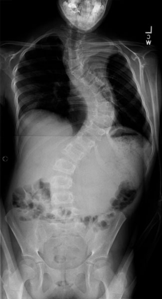 |
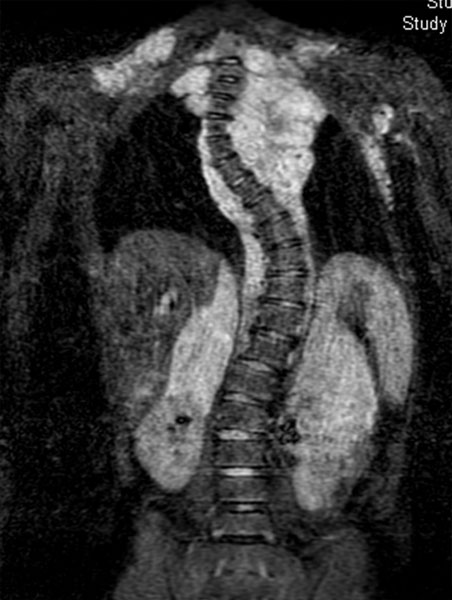 |
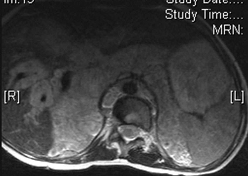 |
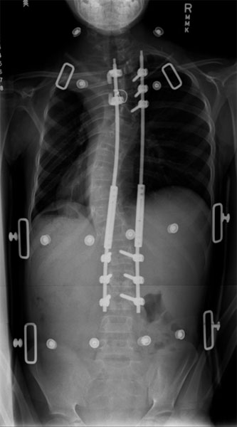 |
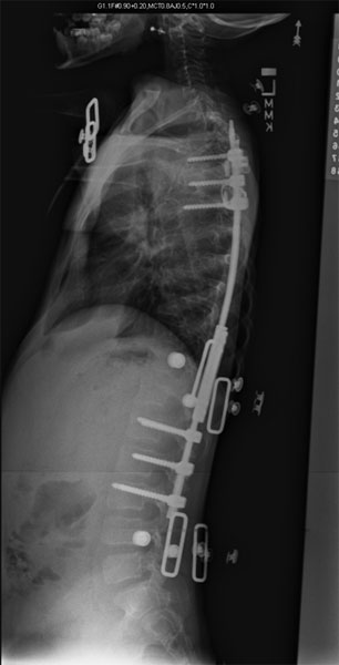 |
The use of growing rod instrumentation in NF1 is also associated with a high incidence of complications (Figure 3). The high rate of complications has also been reported for idiopathic patients.39 The most common complication we have encountered is proximal juctional kyphosis. This is especially common in the patients with high thoracic or cervicothoracic curves. An all screw construct may be less problematic. In these patients, we currently do not perform routine lengthening. Other complications encountered are infection and rod breakage.
Although the use of growing rod instrumentation is associated with higher complication rate, its benefits outweighs the risk in patients with early onset dystrophic scoliosis. Our early results with the use of growing rods remain encouraging. This is a promising technique made especially useful because most dystrophic curves are early onset.
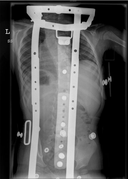 |
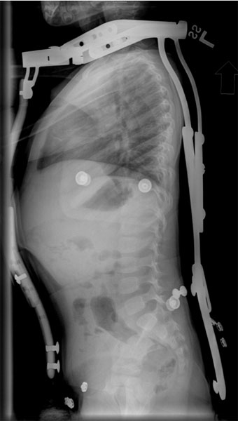 |
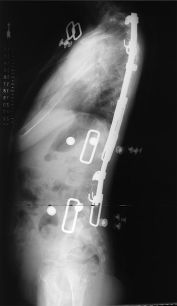 |
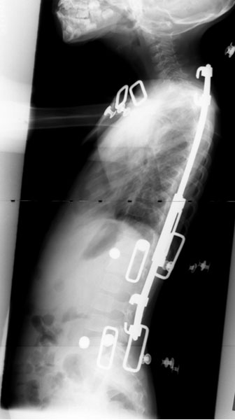 |
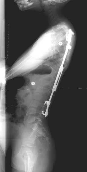 |
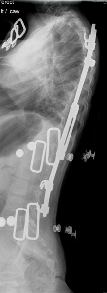 |
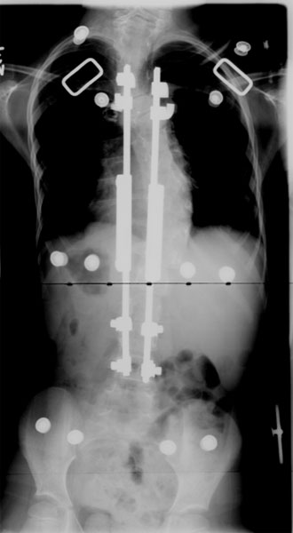 |
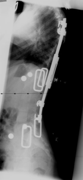 |
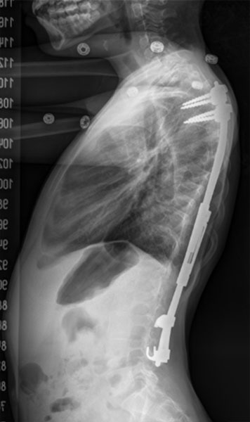 |
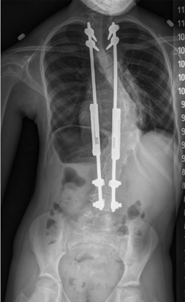 |
Kyphoscoliosis
Kyphoscoliosis seen in NF-1 is distinguished by the predominance of kyphosis (>50 degrees on the lateral radiograph) associated with scoliosis; acute posterior angulation is a typical sign. The vertebral bodies are frequently so deformed and attenuated at the apex that it may be impossible to identify them on routine radiographs.
Patients with dystrophic angular kyphosis respond poorly to posterior fusion alone. This is a potentially aggressive deformity and should be aggressively pursued. Good results are consistently obtained only in those patients who have both anterior and posterior fusion; however, not every patient obtains solid fusion following these procedures. Technically inadequate anterior procedures have been considered the reason for failure; however, pressure erosion from enlarging neurofibromas, dural ectasia, and meningoceles may prevent safe exposure to the front of the spine. Most important, the entire structural area of the deformity should be fused anteriorly with complete disc excision and strong strut grafting, preferably from the fibula, as well as rib and iliac crest graft. No soft tissue should be allowed to interpose between the grafts, and all grafts should have contact with each other and with the spine.
Because of the association of paraplegia with kyphoscoliosis, there has been a tendency to perform laminectomies. Laminectomy alone for kyphoscoliosis cord compression is absolutely contraindicated; the inciting lesion is usually anterior, and the compression cannot be visualized from behind. Also, the removal of the posterior element predisposes the patient to unstable postlaminectomy kyphosis and removes valuable bone stock required for posterior spinal fusion.
The authors strongly recommends anterior and posterior fusion for all dystrophic kyphotic curvatures greater than 40° coronal Cobb angle. If the kyphosis is greater than 50°, a strong anterior strut graft (preferably fibula) should also be performed.
A complete spine MRI or a complete high-volume CT myelogram should be performed prior to surgical treatment. If there is spinal cord impingement, the problem should be approached directly--anteriorly with partial corpectomy for anterior lesions and a hemilaminectomy for posterior ones, both to be followed by anterior and posterior fusions. The patient should be re-imaged six months postsurgery, with augmentation of the fusion mass if there is any evidence of weakness of the fusion mass.
Lordoscoliosis
Lordoscoliosis has been reported in only a small percentage of patients with NF-1. (Figure 4) It is well known that thoracic lordosis (hypokyphosis) predisposes to a significant decrease in pulmonary function and mitral valve prolapse 41, 42
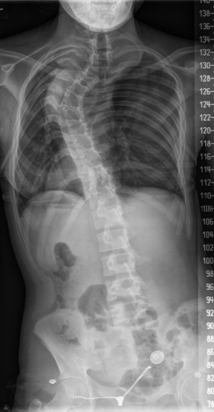 |
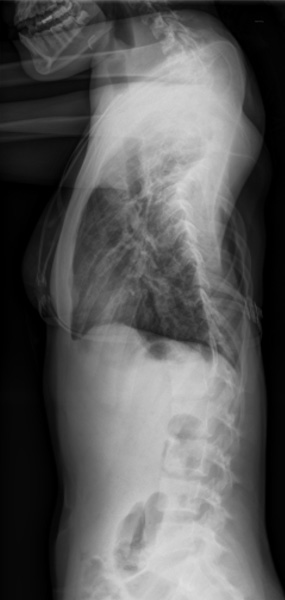 |
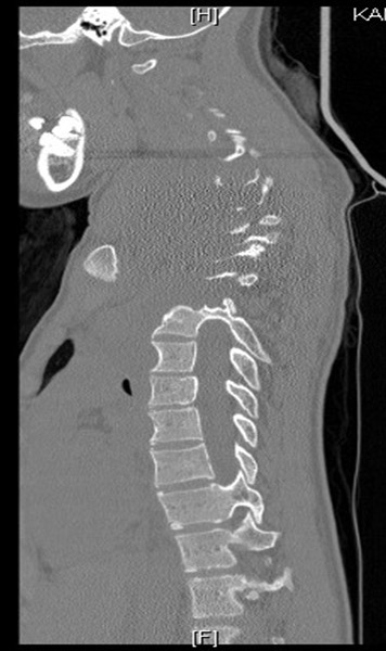 |
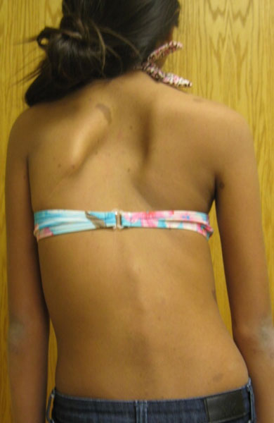 |
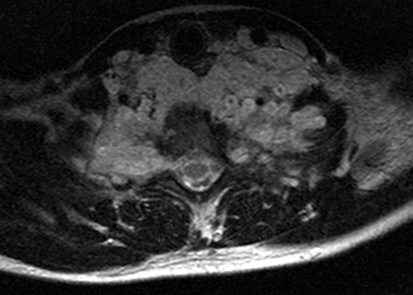 |
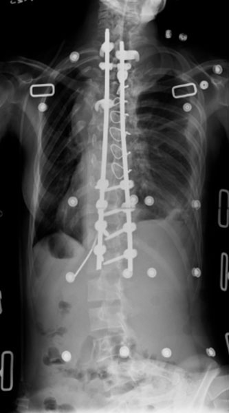 |
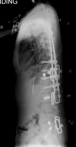 |
Spondylolisthesis
The deformity is usually the result of increased diameter of the spinal canal, with pathologic elongation and thinning of the pedicles giving rise to a pathologic forward progression of the anterior elements of the spinal column. Dural ectasia with meningoceles,or interstitial hypertrophic neurofibromas with involvement of the lumbosacral roots exiting the spinal canal through the neuroforaminae, is the cause of the problem. An MRI or large-volume CT myelogram is necessary to identify the character of the soft tissue affecting and surrounding the vertebral elements before considering surgery. The vertebral bodies are secondarily affected and appear to be thin and dystrophic; the smaller vertebral bodies and narrower pedicles may preclude use with pedicle screws. Every effort should be made to achieve anterior and posterior fusion.
Paraplegia
It is possible that neurologic compromise may be related to tumor, structural instability of the vertebral column complex, dural ectasia, vertebral destruction, neurofibromas, neurosarcomas, fibrofatty tissue reaction, severe kyphosis, vertebral subluxation, dislocation, protrusion of ribs into the spinal canal, or progressive dystrophy of the bony elements of the spine. A neoplasm is usually responsible for paraplegia in older patients, whereas spinal malalignment or ribs displacing into the spinal canal is the most common cause in younger individuals. Rockower et al.43 reported two patients who, because of vertebral body instability and displacement secondary to neurofibromatous tissue encroachment, developed paraplegia. The problem was solved by carefully monitored traction and spinal fusion. Traction should be used very rarely when the deformity is mostly kyphotic. Traction should be used only with flexible kyphosis--never if the kyphosis is rigid. If the kyphosis is rigid, an anterior release, disc excision, and fusion followed by posterior spinal fusion are recommended.44
Unrecognized rib penetration into the spinal canal with spinal cord compromise could be a source of paraplegia that, prior to current imaging technology was thought to be the natural course of NF-1 with kyphoscoliosis.
Those with severe spinal curvatures without significant kyphosis and with evidence of paraplegia should be assumed to have intraspinal lesions until proved otherwise. An MRI or a high-volume CT myelogram is done in the prone, lateral, and supine positions. If the kyphosis is mobile and no intraspinal tumor is present, the patient should be placed in halo-assisted traction; this must be done with extreme caution and neurologic monitoring; it should definitely not be performed if the kyphosis is rigid. Even if the kyphosis is mobile and there is paraplegia, the author recommends somatosensory evoked potential monitoring during traction. If a tumor is anterior, immediate anterior excision, spinal cord decompression, and fusion should be carried out; if the lesion is posterior, a hemilaminectomy with tumor excision and posterior spinal fusion should be performed. If total laminectomy is required, posterior spinal fusion is mandatory.
Problems Related To Soft-tissue Involvement
Dural ectasia is a circumferential dilatation of the dural sac which is filled with proteinaceous fluid. The slow expansion of the dura results in erosion of the surrounding osseous structures resulting in widening of the spinal canal, thinning of the laminae, and ultimately destabilization of the spine. Similar lesions are seen in other connective tissue disorders e.g. Marfan’s syndrome and Ehler Danlos syndrome, although cause of these lesions in NF1 is not known.
During this process, the neural elements are not affected. As a result of slow nature of this process and enormous widening of the spinal canal the neural elements have adequate room for accommodation, and there may be severe angular deformity and distortion without neurological deficit. The patients remain neurologically intact until later in the course of the disease process when destabilization of the vertebral column jeopardizes the neural elements. Dislocation of the vertebral column due to dural ectasia has been reported in the literature.45 The destabilization at the costovertebral junction can result in penetration of the rib head into the spinal canal with neurologic compromise (Figure 1).44, 46 The presence of rib head or the neurofibroma in the spinal canal can result in intra-operative neurological deficit if instrumentation is used for correction of the curve without adequate decompression.
Dural ectasia can be readily seen on high-volume computed tomographic (CT) myelography or contrast-enhanced MR imaging and is recommended before surgical intervention is undertaken for dystrophic curves. Higher imaging studies help to demonstrate extremely thin laminae; in which case dissection by electocautery rather than by periosteal elevators are recommended during surgical exposure to avoid direct injury to the neural elements/dura by plunging into the spinal canal. Surgical spinal stabilization and fusion does not alter the course of dural ectasia. Dural ectasia can result in failure of the primary fusion or the expanding dura ultimately can destroy a solid fusion leaving behind the instrumentation.
Intrathoracic Meningocele
A meningocele is a protrusion of the spinal meninges through an intervertebral foramen or bony defect of the vertebra; it contains an extension of the subarachnoid space filled with cerebrospinal fluid. Meningocele in association with NF-1 can occur at any level of the spine. A posterior mediastinal mass in a patient with NF-1, particularly if associated with kyphoscoliosis, is most likely a lateral meningocele. With MRI, a well-demarcated soft-tissue cystic mass is seen protruding from the spinal canal into the posterior mediastinum. Structural defects in the pedicles, enlargement of the intervertebral foramina, rib deformities including costotransverse dislocation, and scalloping of the vertebral bodies may accompany the advancing mass.
Because intrathoracic meningoceles are often symptomless, the question of treatment is difficult. If it is a chance radiologic finding and causes no symptoms, the right course would be observation. If there is definite progressive enlargement, an initial attempt should be made to ligate the sac. It may be feasible to occlude the neck of the sac by plicating sutures via video-assisted thoracoscopy. Excision of the lesion is indicated for progressive excavation of the vertebra, neurologic injury, respiratory distress or if there is evidence of rapid progression in size.
Dumbbell Lesions
The classic dumbbell lesion is one in which the neurofibroma is constricted as it exits the neuroforamina, giving it the appearance of a weight lifter's dumbbell. (Figure #13A) They may be intradural and extradural, intradural, extradural, or extradural and extraspinal. The tumors may occur at any level of the cord, but the cervical and thoracic levels are most often involved. The tumors may present as nodules arising from the sheath along the nerve, or they may actually invade the nerve. If the tumor invades the nerve with consequent interstitial hypertrophy, then “the nerve becomes the tumor and the tumor becomes the nerve.” And removing the tumor results in a neurologic deficit. The incidence of malignant degeneration of peripheral neurofibromas is unknown.
Thoracic Tumors
We recently reviewed 260 pediatric patients with NF-1 and noted nine patients (3.5%) to have extrapleural thoracic tumors. Ninety-five of these patients were primarily screened by chest x-rays, and many patients might have asymptomatic thoracic tumors that have yet to be diagnosed. All patients were 6 years or older, six were asymptomatic, three presented with respiratory symptoms. A clue to thoracic involvement in two patients was the presence of a visible plexiform neurofibroma in the neck, which later was found to have extended into the chest. Focal scoliosis was a significant clue to an underlying malignant transformation of a benign plexiform neurofibroma occurred in one of our patients.
Signs on physical examination that should raise suspicion of an underlying thoracic tumor include a plexiform neurofibroma of the neck or a focal area of scoliosis. When reviewed retrospectively, eight of the nine patients had either a symptom or a sign on physical examination that was a clue to the underlying tumor. Surgical management of these tumors can be difficult because of frequent involvement of nerves and blood vessels.47
Vertebral Column Dislocation
Complete dislocation of the spine in NF-1 is rare. Significant subluxation or dislocation of the spine in patients who have NF-1 can occur with little radiographic or clinical warning, because the osseous erosion is so extensive. This diagnosis should be considered in any patient who has NF-1 and unexplained pain in the neck or back.
Plexiform Venous Channels
Plexiform venous anomalies may surround the spine, impeding the operative approach to the vertebral bodies. They may be so dense that the anterior approach to the internal kyphosis had to be abandoned because of excessive bleeding (34). The authors have not experienced excessive venous bleeding while performing anterior removal of neurofibromatous tissue and strut grafting, but warns the reader of the possibility. Wound hematomas following surgery have not been a problem; however, meticulous hemostasis and wound drainage should be carried out when performing surgery on patients with NF-1.
References
1. Fienman NL. Pediatric neurofibromatosis: review. Compr Ther 1981;7:66-72.
2. Crawford AH, Schorry EK. Neurofibromatosis update. J Pediatr Orthop 2006;26:413-23.
3. Calzavara PG, Carlino A, Anzola GP, Pasolini MP. Segmental neurofibromatosis. Case report and review of the literature. Neurofibromatosis 1988;1:318-22.
4. Kaufman DL, Heinrich BS, Willett C, et al. Somatic instability of the NF2 gene in schwannomatosis. Arch Neurol 2003;60:1317-20.
5. National Institutes of Health Consensus Development Conference Statement: neurofibromatosis. Bethesda, Md., USA, July 13-15, 1987. Neurofibromatosis 1988;1:172-8.
6. Crawford AH, Schorry EK. Neurofibromatosis in children: the role of the orthopaedist. J Am Acad Orthop Surg 1999;7:217-30.
7. Listernick R, Charrow J. Neurofibromatosis type 1 in childhood. J Pediatr 1990;116:845-53.
8. Stevenson DA, Viskochil DH, Schorry EK, et al. The use of anterolateral bowing of the lower leg in the diagnostic criteria for neurofibromatosis type 1. Genet Med 2007;9:409-12.
9. Collins FS, Ponder BA, Seizinger BR, Epstein CJ. The von Recklinghausen neurofibromatosis region on chromosome 17--genetic and physical maps come into focus. Am J Hum Genet 1989;44:1-5.
10. Goldberg NS, Collins FS. The hunt for the neurofibromatosis gene. Arch Dermatol 1991;127:1705-7.
11. Korf BR. The phakomatoses. Clin Dermatol 2005;23:78-84.
12. Messiaen LM, Callens T, Mortier G, et al. Exhaustive mutation analysis of the NF1 gene allows identification of 95% of mutations and reveals a high frequency of unusual splicing defects. Hum Mutat 2000;15:541-55.
13. Crawford AH. Neurofibromatosis. 3 ed. Philadelphia: Lippincott; 1990.
14. Akbarnia BA, Gabriel KR, Beckman E, Chalk D. Prevalence of scoliosis in neurofibromatosis. Spine 1992;17:S244-8.
15. Halmai V, Doman I, de Jonge T, Illes T. Surgical treatment of spinal deformities associated with neurofibromatosis type 1. Report of 16 cases. Journal of neurosurgery 2002;97:310-6.
17. Durrani AA, Crawford AH, Chouhdry SN, Saifuddin A, Morley TR. Modulation of spinal deformities in patients with neurofibromatosis type 1. Spine 2000;25:69-75.
18. Shufflebarger HL. Cotrel-Dubousset instrumentation in neurofibromatosis spinal problems. Clinical orthopaedics and related research 1989:24-8.
19. Betz RR, Iorio R, Lombardi AV, Clancy M, Steel HH. Scoliosis surgery in neurofibromatosis. Clinical orthopaedics and related research 1989:53-6.
20. Hunt JC, Pugh DG. Skeletal lesions in neurofibromatosis. Radiology 1961;76:1-20.
21. Crawford AH. Neurofibromatosis in children. Acta orthopaedica Scandinavica 1986;218:1-60.
22. Crawford AH. Neurofibromatosis. In: Weinstein SL, ed. The pediatric spine: Principles and practice. New York: Raven Press; 1994:619-49.
23. Winter RB, Moe JH, Bradford DS, Lonstein JE, Pedras CV, Weber AH. Spine deformity in neurofibromatosis. A review of one hundred and two patients. The Journal of bone and joint surgery 1979;61:677-94.
23. Crawford AH. Pitfalls of spinal deformities associated with neurofibromatosis in children. Clinical orthopaedics and related research 1989:29-42.
24. Kim HW, Weinstein SL. Spine update. The management of scoliosis in neurofibromatosis. Spine 1997;22:2770-6.
25. Funasaki H, Winter RB, Lonstein JB, Denis F. Pathophysiology of spinal deformities in neurofibromatosis. An analysis of seventy-one patients who had curves associated with dystrophic changes. The Journal of bone and joint surgery 1994;76:692-700.
26. Holt RT, Johnson JR. Cotrel-Dubousset instrumentation in neurofibromatosis spine curves. A preliminary report. Clinical orthopaedics and related research 1989:19-23.
27. Sirois JL, 3rd, Drennan JC. Dystrophic spinal deformity in neurofibromatosis. J Pediatr Orthop 1990;10:522-6.
28. Crawford AH, Parikh S, Schorry EK, Von Stein D. The immature spine in type-1 neurofibromatosis. The Journal of bone and joint surgery 2007;89 Suppl 1:123-42.
29. Crawford AH. Neurofibromatosis. In: Weinstein SL, ed. The pediatric spine: Principles and practice. Philadelphia: Lippincott Williams & Wilkins; 2001:471-90.
30. Calvert PT, Edgar MA, Webb PJ. Scoliosis in neurofibromatosis. The natural history with and without operation. The Journal of bone and joint surgery 1989;71:246-51.
31. Mehlman CT, Al-Sayyad MJ, Crawford AH. Effectiveness of spinal release and halo-femoral traction in the management of severe spinal deformity. J Pediatr Orthop 2004;24:667-73.
32. Rinella A, Lenke L, Whitaker C, et al. Perioperative halo-gravity traction in the treatment of severe scoliosis and kyphosis. Spine 2005;30:475-82.
33. Winter RB, Lonstein JE, Anderson M. Neurofibromatosis hyperkyphosis: a review of 33 patients with kyphosis of 80 degrees or greater. Journal of spinal disorders 1988;1:39-49.
34. Lalueza MP, Colomina MJ, Bago J, Clemente S, Godet C. Analysis of nutritional parameters in idiopathic scoliosis patients after major spinal surgery. European journal of clinical nutrition 2005;59:720-2.
35. Al-Sayyad MJ, Crawford AH, Wolf RK. Early experiences with video-assisted thoracoscopic surgery: our first 70 cases. Spine 2004;29:1945-51; discussion 52.
36. Mulpuri K, LeBlanc JG, Reilly CW, et al. Sternal split approach to the cervicothoracic junction in children. Spine 2005;30:E305-10.
37. Parisini P, Di Silvestre M, Greggi T, Paderni S, Cervellati S, Savini R. Surgical correction of dystrophic spinal curves in neurofibromatosis. A review of 56 patients. Spine 1999;24:2247-53.
38. Singh K, Samartzis D, An HS. Neurofibromatosis type I with severe dystrophic kyphoscoliosis and its operative management via a simultaneous anterior-posterior approach: a case report and review of the literature. Spine J 2005;5:461-6.
39. Akbarnia BA, Marks DS, Boachie-Adjei O, Thompson AG, Asher MA. Dual growing rod technique for the treatment of progressive early-onset scoliosis: a multicenter study. Spine 2005;30:S46-57.
40. Thompson GH, Akbarnia BA, Kostial P, et al. Comparison of single and dual growing rod techniques followed through definitive surgery: a preliminary study. Spine 2005;30:2039-44.
41. Hirschfeld SS, Rudner C, Nash CL, Jr., Nussbaum E, Brower EM. Incidence of mitral valve prolapse in adolescent scoliosis and thoracic hypokyphosis. Pediatrics 1982;70:451-4.
42. Winter RB, Lovell WW, Moe JH. Excessive thoracic lordosis and loss of pulmonary function in patients with idiopathic scoliosis. The Journal of bone and joint surgery 1975;57:972-7.
43. Rockower S, McKay D, Nason S. Dislocation of the spine in neurofibromatosis. A report of two cases. The Journal of bone and joint surgery 1982;64:1240-2.
44. Major MR, Huizenga BA. Spinal cord compression by displaced ribs in neurofibromatosis. A report of three cases. The Journal of bone and joint surgery 1988;70:1100-2.
45. Stone JW, Bridwell KH, Shackelford GD, Abramson CL. Dural ectasia associated with spontaneous dislocation of the upper part of the thoracic spine in neurofibromatosis. A case report and review of the literature. The Journal of bone and joint surgery 1987;69:1079-83.
46. Flood BM, Butt WP, Dickson RA. Rib penetration of the intervertebral foraminae in neurofibromatosis. Spine 1986;11:172-4.
47. Schorry EK, Crawford AH, Egelhoff JC, Lovell AM, Saal HM. Thoracic tumors in children with neurofibromatosis-1. American journal of medical genetics 1997;74:533-7.